Skeletal X-Rays for Bone Fracture Diagnosis: Comprehensive Imaging and Patient Guidance
Skeletal X-rays are a vital tool in diagnosing bone fractures and injuries, providing clear images that help medical professionals assess the extent of damage. This article will explore the various types of bone fractures diagnosed through X-rays, the process of interpreting these images, and how patients can prepare for their appointments. Understanding the role of skeletal X-rays is crucial for anyone seeking effective diagnosis and treatment for bone-related injuries. We will also discuss advanced imaging techniques, specialized services, and the importance of bone health awareness in South Africa.
What Are the Common Types of Bone Fractures Diagnosed by X-Ray?
Bone fractures can be classified into several types, each with distinct characteristics visible on X-rays. Understanding these types is essential for accurate diagnosis and treatment planning.
How Do Transverse, Oblique, and Spiral Fractures Appear on Skeletal X-Rays?
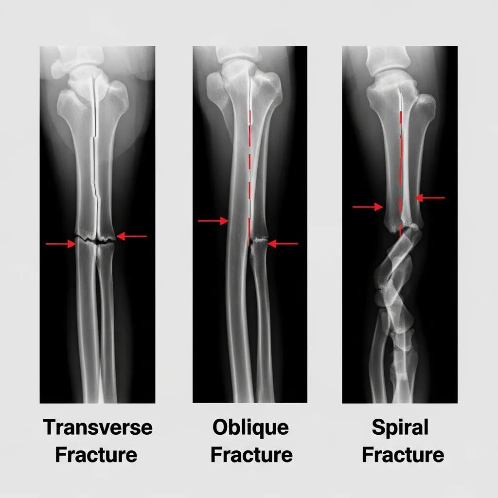
Transverse fractures are characterized by a straight break across the bone, often resulting from a direct impact. Oblique fractures, on the other hand, occur at an angle and are typically caused by a twisting force. Spiral fractures are more complex, resulting from a rotational force that causes the bone to break in a spiral pattern. Each type of fracture has a unique appearance on X-rays, which aids in determining the appropriate treatment approach.
What Are Pediatric Fractures and How Are Greenstick and Growth Plate Injuries Detected?
Pediatric fractures often differ from adult fractures due to the flexibility of children’s bones. Greenstick fractures, for instance, are incomplete fractures where the bone bends and cracks on one side without breaking completely. Growth plate injuries are critical as they can affect a child’s future bone growth. X-rays are essential in detecting these injuries, allowing for timely intervention to prevent long-term complications.
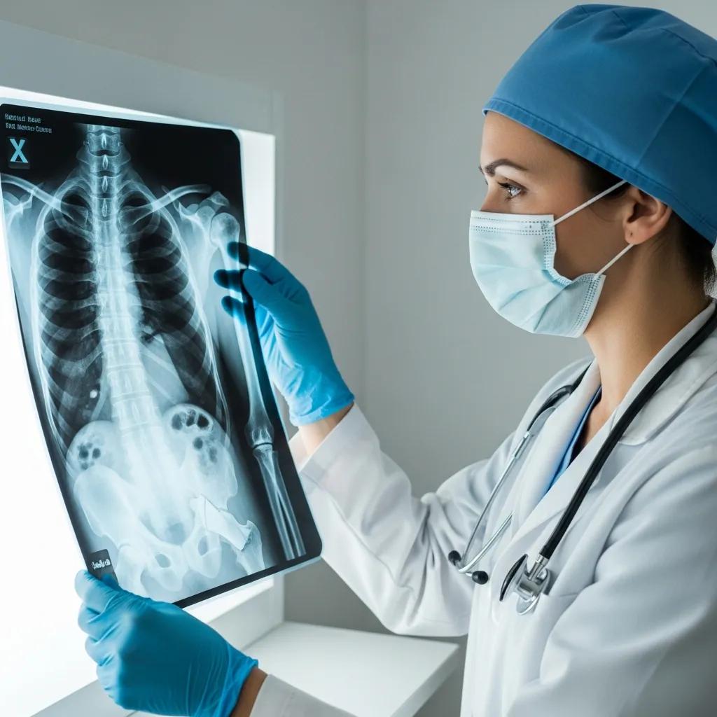
How Do Skeletal X-Rays Detect Bone Fractures and Injuries?
Skeletal X-rays are a primary diagnostic tool for identifying bone fractures and injuries. They provide detailed images that help radiologists and physicians assess the condition of bones.
What Is the Process of Interpreting a Skeletal X-Ray Report?
Interpreting a skeletal X-ray report involves analyzing the images for signs of fractures, misalignments, or other abnormalities. Radiologists look for specific indicators, such as the fracture line’s location and the surrounding bone’s condition. Understanding the terminology used in reports is crucial for both healthcare providers and patients to ensure accurate communication regarding the diagnosis.
What Are the Benefits and Limitations of X-Rays in Diagnosing Bone Injuries?
X-rays offer several benefits in diagnosing bone injuries, including their ability to provide immediate results and their widespread availability. However, they also have limitations, such as their inability to visualize soft tissues and certain complex fractures. In some cases, additional imaging techniques like CT scans or MRIs may be necessary for a comprehensive assessment.
When to Use CT and MRI for Detailed Skeletal Injury Assessment
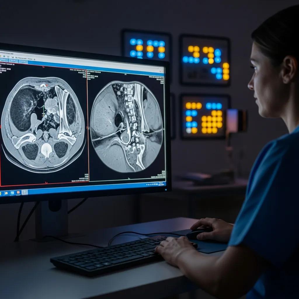
While X-rays are effective for many fractures, advanced imaging techniques may be required for a more detailed evaluation of complex injuries.
How Do CT Scans Assist in Assessing Complex Bone Fractures?
CT scans provide cross-sectional images of the body, allowing for a more detailed view of complex bone fractures. They are particularly useful in assessing fractures involving joints or those that are difficult to visualize with standard X-rays. This advanced imaging technique helps in planning surgical interventions and ensuring optimal treatment outcomes.
What Role Does MRI Play in Detecting Soft Tissue Injuries Associated with Bone Trauma?
MRI is invaluable for detecting soft tissue injuries that may accompany bone trauma, such as ligament tears or muscle damage. Unlike X-rays, MRIs provide detailed images of both bone and soft tissue, making them essential for a comprehensive evaluation of skeletal injuries. This capability is crucial for developing effective treatment plans that address all aspects of an injury.
How Can Patients Prepare for Skeletal X-Ray Appointments at Tesla Radiological Services?
Preparing for a skeletal X-ray appointment can help ensure a smooth and efficient process. Patients should be aware of what to expect during their visit.
What Should Patients Expect During Their Bone Fracture X-Ray Procedure?
During a bone fracture X-ray procedure, patients will be positioned to capture the necessary images of the affected area. The process is quick, typically taking only a few minutes. Patients may need to remove any clothing or accessories that could interfere with the imaging. Understanding these steps can help alleviate any anxiety about the procedure.
How Does Tesla Radiological Services Facilitate Convenient Online Booking and Multiple Locations?
Tesla Radiological Services offers convenient online booking options, allowing patients to schedule their appointments easily. With multiple locations available, patients can choose a facility that best suits their needs, ensuring accessibility and convenience for all. This streamlined process enhances the overall patient experience, making it easier to receive timely care.
What Specialized Skeletal Imaging Services Are Available for Specific Patient Needs?
Specialized skeletal imaging services cater to various patient needs, ensuring comprehensive care for different conditions.
How Are Sports Injuries and Pediatric Skeletal Conditions Diagnosed with X-Rays?
Sports injuries often require specialized imaging to assess the extent of damage. X-rays are commonly used to diagnose fractures and other injuries in athletes, allowing for prompt treatment. Similarly, pediatric skeletal conditions, such as growth plate injuries, are effectively diagnosed using X-rays, ensuring that young patients receive the appropriate care.
What Imaging Services Support Workmen's Compensation and Road Accident Fund Claims?
Imaging services play a crucial role in supporting workmen’s compensation and road accident fund claims. Accurate X-ray imaging is essential for documenting injuries sustained in accidents, providing the necessary evidence for claims processing. This service ensures that patients receive the support they need during recovery.
How Does Skeletal Imaging Support Bone Health Awareness and Fracture Prevention in South Africa?
Skeletal imaging is vital for promoting bone health awareness and preventing fractures, particularly in populations at risk.
What Is the Importance of Bone Density Scans in Osteoporosis Diagnosis?
Bone density scans are essential for diagnosing osteoporosis, a condition that weakens bones and increases fracture risk. These scans help identify individuals at risk, allowing for early intervention and preventive measures. Understanding the importance of bone health is crucial for reducing the incidence of fractures in the community.
How Is Tesla Radiological Services Addressing the Growing Fracture Burden and Radiologist Shortage?
Tesla Radiological Services is actively addressing the growing fracture burden and the shortage of radiologists by implementing innovative solutions. By enhancing imaging capabilities and streamlining processes, the organization aims to provide timely and accurate diagnoses, ensuring that patients receive the care they need without unnecessary delays. This commitment to improving skeletal imaging services is vital for supporting public health in South Africa.