Digital X-Ray vs Traditional X-Ray: Understanding Key Differences and Benefits
In the realm of medical imaging, the evolution from traditional x-ray methods to digital x-ray technology marks a significant advancement in diagnostic capabilities. This article delves into the key differences between digital x-ray and traditional x-ray, highlighting the mechanisms, benefits, and technological innovations that define each method. Readers will gain insights into how these imaging techniques operate, their respective advantages, and the implications for patient safety and diagnostic accuracy. As healthcare continues to embrace technological advancements, understanding these differences is crucial for both practitioners and patients alike. We will explore the operational processes, technological advancements, and the overall impact on patient care, culminating in how patients can access digital x-ray services.
Core Differences Between Digital and Traditional X-Ray Systems
Digital x-ray and traditional x-ray imaging differ fundamentally in their technology and processes. Traditional x-ray imaging utilizes film to capture images, which requires chemical processing and can take time to develop. In contrast, digital x-ray employs electronic sensors to capture images instantly, allowing for immediate viewing and analysis. This shift not only enhances efficiency but also reduces the need for physical storage of films, streamlining the workflow in medical facilities.
The key differences can be summarized as follows:
- Image Capture: Traditional x-rays use film, while digital x-rays use electronic sensors.
- Processing Time: Digital x-rays provide immediate results, whereas traditional methods require time for film development.
- Storage and Accessibility: Digital images can be stored electronically, making them easier to access and share compared to physical films.
Understanding these differences is essential for evaluating the benefits of each method in clinical practice.
How Does Digital Radiography Work Compared to Traditional X-Ray Processes?
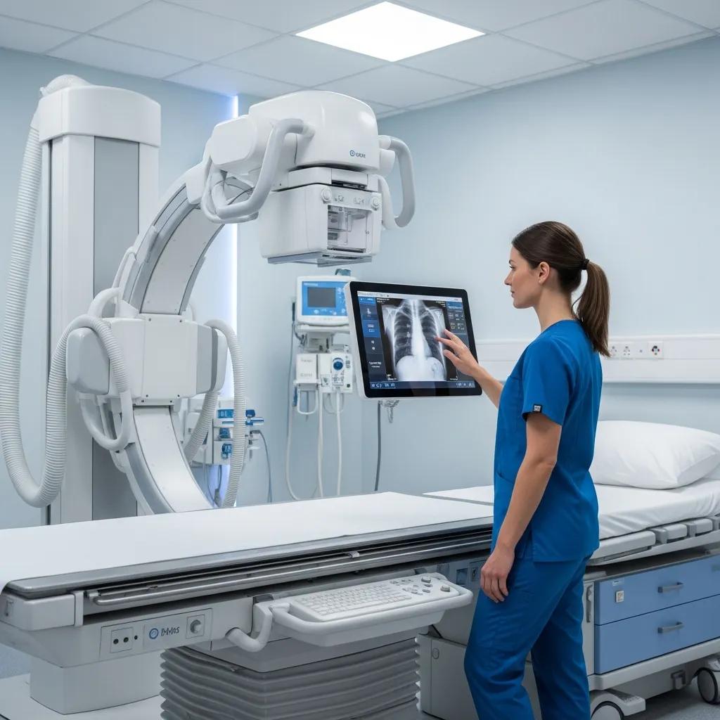
Digital radiography operates by using digital sensors that convert x-ray energy into electronic signals, which are then processed to create images. This process is significantly faster than traditional x-ray methods, where images are captured on film and require chemical processing. The digital approach allows for real-time image viewing, enabling healthcare providers to make quicker diagnostic decisions.
In traditional x-ray processes, the film is exposed to x-rays, and the resulting image is developed using chemical solutions. This method not only takes longer but also involves additional steps that can introduce variability in image quality. Digital radiography eliminates these steps, resulting in a more efficient and reliable imaging process.
Further emphasizing the efficiency gains, studies highlight how digital radiography significantly streamlines clinical operations and patient flow.
Digital Radiography: Enhancing Clinical Workflow & Throughput
It is commonly accepted that digital radiography (DR) improves workflow and patient throughput compared with traditional film radiography or computed radiography (CR). DR eliminates the film development step and the time to acquire the image from a CR reader. In addition, the wide dynamic range of DR is such that the technologist can perform the quality-control (QC) step directly at the modality in a few seconds, rather than having to transport the newly acquired image to a centralized QC station for review. Furthermore, additional workflow efficiencies can be achieved with DR by employing tight radiology information system (RIS) integration.
Impact of digital radiography on clinical workflow, 2000
What Are the Technological Advances in Digital X-Ray Machines?
Recent advancements in digital x-ray technology have led to improved image quality and diagnostic capabilities. Some notable technological innovations include:
- Higher Resolution Sensors: Modern digital x-ray machines utilize advanced sensors that provide clearer and more detailed images, enhancing diagnostic accuracy.
- Image Enhancement Software: Software tools allow for image manipulation, enabling radiologists to adjust contrast and brightness for better visibility of structures.
- Wireless Technology: Many digital x-ray systems now feature wireless capabilities, facilitating easier image transfer and reducing clutter in examination rooms.
These advancements not only improve the quality of images but also enhance the overall efficiency of the diagnostic process.
What Are the Advantages of Digital X-Ray Over Traditional Methods?
Digital x-ray technology offers several advantages over traditional x-ray methods, making it a preferred choice in many medical facilities. The primary benefits include:
| Advantage | Description |
|---|---|
| Reduced Radiation Exposure | Digital x-rays require less radiation to produce high-quality images, minimizing patient exposure. |
| Faster Imaging Results | Images are available for review almost instantly, allowing for quicker diagnosis and treatment. |
| Enhanced Diagnostic Accuracy | The ability to manipulate images digitally improves the clarity and detail, aiding in more accurate diagnoses. |
These advantages contribute to better patient outcomes and more efficient healthcare delivery.
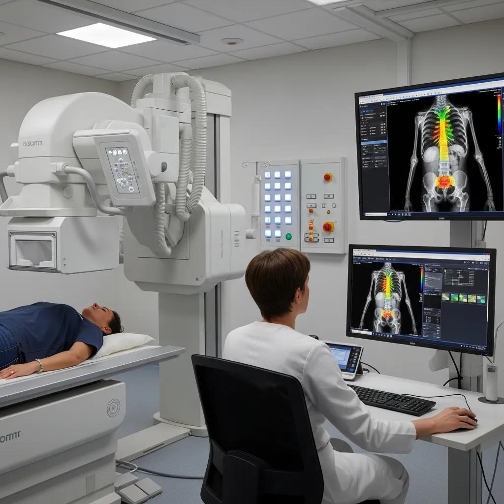
How Does Digital X-Ray Improve Diagnostic Accuracy and Image Quality?
Digital x-ray technology enhances diagnostic accuracy through several mechanisms. The high-resolution images produced by digital sensors allow for better visualization of anatomical structures, which is crucial for accurate diagnosis. Additionally, the use of image enhancement software enables radiologists to adjust images for optimal clarity, making it easier to identify abnormalities.
Furthermore, digital x-rays can be easily compared with previous images, allowing for more effective monitoring of changes over time. This capability is particularly beneficial in tracking the progression of diseases or the healing process after treatment.
The continuous development of digital imaging technology, including advanced noise reduction algorithms, further contributes to both image quality and patient safety at lower doses.
Digital X-Ray: Dose Reduction & Image Quality with Noise Algorithms
PurposeTo evaluate the image quality of low-dose chest digital radiographic images obtained with a new spatial noise reduction algorithm, compared to a conventional de-noising technique.Materials and methodsIn 69 patients, the dose reduction protocol was divided into A, B, and C test groups– 60% (n = 22), 50% (n = 23), and 40% (n = 24) of the baseline dose. In each patient, baseline dose radiographs were obtained with conventional image processing while low-dose images were acquired with new image processing.
Radiation dose reduction and improvement of image quality in digital chest radiography by new spatial noise reduction algorithm, W Lee, 2020
In What Ways Does Digital Radiography Enhance Patient Safety and Speed?
Digital radiography significantly enhances patient safety by reducing radiation exposure and improving the speed of imaging processes. The lower doses of radiation required for digital x-rays minimize the risk associated with imaging procedures. Additionally, the rapid availability of images allows healthcare providers to make timely decisions regarding patient care, which is critical in emergency situations.
Moreover, the streamlined workflow associated with digital imaging reduces the time patients spend in the imaging department, contributing to a more efficient overall experience.
How Does Radiation Exposure Compare Between Digital and Traditional X-Rays?
When comparing radiation exposure between digital and traditional x-rays, digital x-rays consistently demonstrate lower exposure levels. The advanced technology used in digital imaging allows for high-quality images to be captured with significantly less radiation. This reduction in exposure is particularly important for vulnerable populations, such as children and pregnant women.
Research indicates that digital x-rays can reduce radiation doses by up to 80% compared to traditional film-based x-rays, making them a safer option for patients.
What Is the Radiation Dose Difference Between Digital and Film-Based X-Rays?
The radiation dose difference between digital and film-based x-rays is substantial. Digital x-ray systems are designed to optimize image quality while minimizing radiation exposure. Studies have shown that digital x-rays can achieve the same diagnostic quality as traditional x-rays with much lower radiation doses.
For example, a typical digital x-ray may require only 0.1 mSv of radiation, while a traditional film x-ray might require 0.2 mSv or more. This significant difference underscores the safety benefits of digital imaging technologies.
Indeed, advanced noise reduction technologies have been shown to dramatically decrease patient radiation exposure across various procedures.
X-Ray Noise Reduction Technology for Patient Dose Safety
This retrospective study quantifies the patient dose reduction due to the introduction of a novel X-ray imaging noise reduction technology using advanced real-time image noise reduction algorithms and optimized acquisition chain for fluoroscopy and exposure in interventional cardiology. The new system provides significant dose reduction compared to the reference system. Median DAP values decreased for all procedures (p< 0.0001) from 172.7 to 59.4 Gy cm2, for CAG from 155.1 to 52.0 Gy cm2and for PCI from 229.0 to 85.8 Gy cm2with reduction quantified at 66, 66 and 63 %, respectively.
Patient radiation dose reduction using an X-ray imaging noise reduction technology for cardiac angiography and intervention, 2016
Why Is Digital X-Ray Considered Safer for Patients?
Digital x-ray is considered safer for patients due to its lower radiation exposure and the ability to quickly identify and address potential issues. The immediate availability of images allows healthcare providers to act swiftly, reducing the time patients are exposed to radiation. Additionally, the enhanced image quality aids in accurate diagnoses, which can prevent unnecessary repeat imaging.
The integration of digital x-ray technology into clinical practice represents a commitment to patient safety and improved healthcare outcomes.
How Can Patients Book Digital X-Ray Services with X-Ray Docs?
Patients interested in accessing digital x-ray services can easily book appointments with X-Ray Docs, a leading imaging service provider in South Africa. The process is straightforward and designed to accommodate patient needs.
To book a digital x-ray appointment, patients can visit the X-Ray Docs website or contact their office directly. It is advisable to have relevant medical information and any necessary referrals ready when scheduling an appointment.
What Is the Appointment Booking Process for Digital Radiography at X-Ray Docs?
The appointment booking process for digital radiography at X-Ray Docs involves several simple steps:
- Visit the Website: Navigate to the X-Ray Docs website to access the appointment scheduling section.
- Provide Information: Fill out the required information, including personal details and the reason for the x-ray.
- Select a Date and Time: Choose a convenient date and time for the appointment.
- Confirmation: Receive confirmation of the appointment via email or phone.
This streamlined process ensures that patients can easily access the digital x-ray services they need.
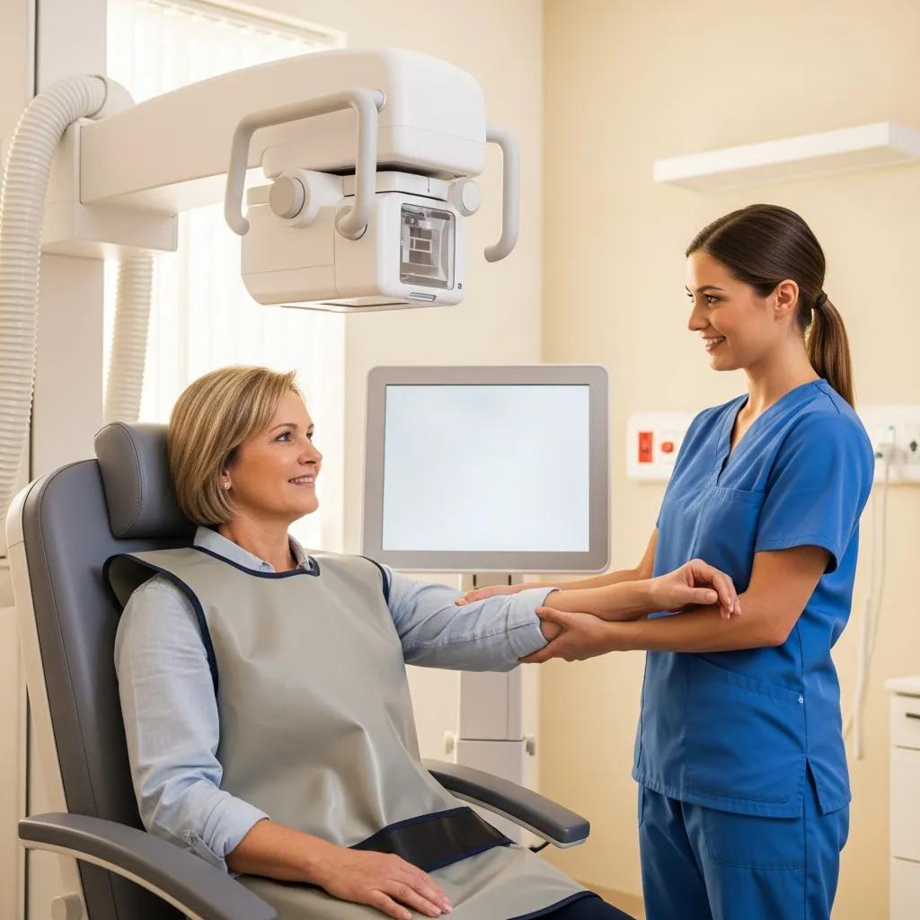
What Should Patients Expect During a Digital X-Ray Procedure?
During a digital x-ray procedure, patients can expect a quick and efficient experience. The process typically involves the following steps:
- Preparation: Patients may be asked to remove any clothing or accessories that could interfere with the imaging.
- Positioning: A technician will position the patient appropriately to capture the necessary images.
- Imaging: The digital x-ray machine will take the images, which are available for immediate review.
Patients often find the process to be comfortable and less time-consuming compared to traditional x-ray methods. The use of digital technology enhances the overall experience, making it a preferred choice for many.