Shoulder Injury Imaging: Comparing MRI and X-Ray Accuracy for Rotator Cuff Tear Diagnosis
Shoulder injuries, particularly rotator cuff tears, are common and can significantly impact daily activities. Accurate imaging is crucial for effective diagnosis and treatment. This article explores the differences between MRI and X-ray imaging techniques, focusing on their effectiveness, advantages, and limitations in diagnosing rotator cuff tears. Readers will learn about the key symptoms that necessitate imaging, the anatomy of the rotator cuff, and how these factors influence the choice of imaging modality. Additionally, we will discuss how MRI facilitates accurate diagnosis and the procedure involved, while also addressing the role of X-ray in assessing shoulder injuries. Finally, we will provide guidance on how patients can choose between these imaging options and book appointments at X-Ray Docs.
Effectiveness:
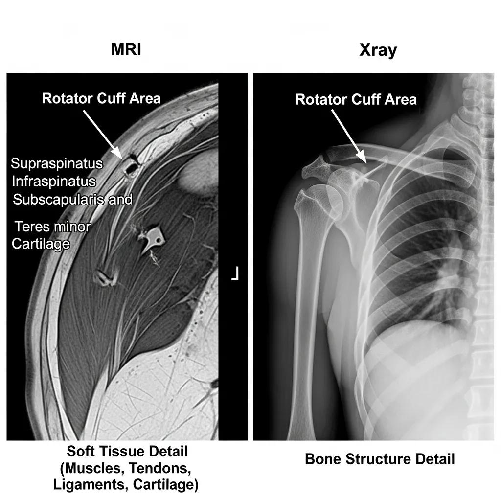
MRI and X-ray are both valuable tools in diagnosing rotator cuff tears, but they serve different purposes. MRI is particularly effective in providing detailed images of soft tissues, including muscles, tendons, and ligaments, which are crucial for identifying rotator cuff injuries. In contrast, X-ray primarily visualizes bone structures, making it less effective for soft tissue assessment. Understanding the effectiveness of these imaging modalities is essential for accurate diagnosis and treatment planning.
Advantages:
Both MRI and X-ray have distinct advantages that make them suitable for different diagnostic scenarios.
- MRI Advantages: MRI has no radiation exposure, making it a safer option for patients, especially those requiring multiple scans.It provides high-resolution images of soft tissues, allowing for a comprehensive evaluation of rotator cuff injuries.
- X-Ray Advantages: X-ray is generally less expensive and widely available, making it a more accessible option for initial assessments.It is quick to perform, providing immediate results that can guide further diagnostic steps.
These advantages highlight the importance of selecting the appropriate imaging technique based on the clinical context.
Limitations:
While MRI and X-ray are effective, they also have limitations that must be considered.
- MRI Limitations: MRI scans typically have longer scan times, which can be uncomfortable for some patients.The cost of MRI can be significantly higher than that of X-ray, which may limit access for some individuals.
- X-Ray Limitations: X-ray cannot visualize soft tissues, making it inadequate for diagnosing rotator cuff tears.It is primarily useful for assessing bone injuries, such as fractures, rather than soft tissue damage.
Understanding these limitations is crucial for healthcare providers when determining the best imaging approach for shoulder injuries.
What Are the Key Symptoms and Diagnostic Needs for Rotator Cuff Tears?
Key symptoms of rotator cuff tears include persistent shoulder pain, weakness in arm movement, and difficulty performing overhead activities. These symptoms often prompt healthcare providers to recommend imaging studies to confirm the diagnosis. MRI is preferred for detailed imaging of soft tissues, allowing for accurate assessment of the rotator cuff and surrounding structures. Recognizing these symptoms and understanding the diagnostic needs can lead to timely and effective treatment.
How Does Rotator Cuff Anatomy Influence Injury Detection?
The rotator cuff consists of four muscles and their associated tendons, which stabilize the shoulder joint and facilitate movement. Understanding the anatomy of the rotator cuff is essential for detecting injuries. The intricate arrangement of tendons and ligaments can make it challenging to identify tears without advanced imaging techniques. MRI provides a detailed view of these structures, enhancing the ability to detect subtle injuries that may not be visible on X-ray.
Which Clinical Signs Indicate the Need for Imaging?
Several clinical signs indicate the need for imaging in suspected rotator cuff injuries:
- Persistent Pain: Ongoing shoulder pain that does not improve with conservative treatment.
- Weakness: Noticeable weakness in arm movement, particularly when lifting or reaching overhead.
- Limited Range of Motion: Difficulty in moving the shoulder through its full range of motion.
These signs often prompt healthcare providers to recommend imaging studies to confirm the diagnosis and guide treatment.
How Does MRI Imaging Facilitate Accurate Rotator Cuff Tear Diagnosis?
MRI imaging facilitates accurate diagnosis of rotator cuff tears by providing high-resolution images that reveal the condition of soft tissues. The detailed images produced by MRI allow for the identification of tears, inflammation, and other abnormalities in the rotator cuff. Additionally, MRI has a sensitivity of over 90%, which is crucial for ensuring that injuries are not missed. This high sensitivity leads to better diagnosis and timely treatment, ultimately improving patient outcomes.
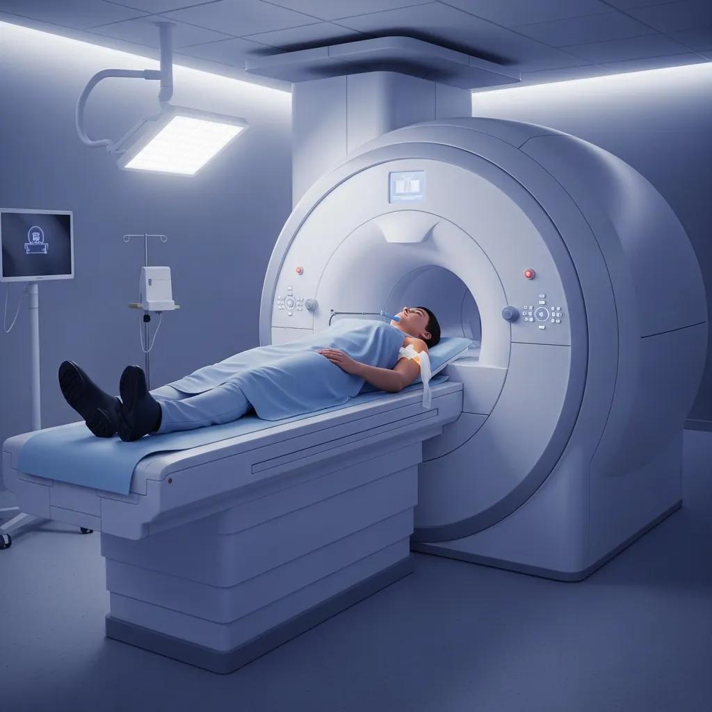
What Is the MRI Procedure for Shoulder Injury Imaging?
The MRI procedure for shoulder injury imaging involves several steps. First, the patient is positioned on the MRI table, and the shoulder is placed within the MRI machine. The procedure uses magnets and radio waves to create detailed images of the shoulder structures. Patients may be required to remain still during the scan, which typically lasts between 20 to 45 minutes. After the scan, a radiologist interprets the images and provides a report to the referring physician.
Why Is MRI Sensitivity Over 90% Important for Rotator Cuff Tears?
The high sensitivity of MRI, exceeding 90%, is critical for diagnosing rotator cuff tears. This level of sensitivity ensures that most injuries are detected, allowing for appropriate treatment to be initiated promptly. Timely diagnosis and intervention can significantly improve patient outcomes, reducing the risk of chronic pain and functional limitations. Understanding the importance of MRI sensitivity helps patients and healthcare providers make informed decisions regarding imaging options.
What Are the Limitations and Uses of X-Ray Imaging for Shoulder Injuries?
X-ray imaging has specific limitations and uses in the context of shoulder injuries. While X-rays are effective for visualizing bone abnormalities, they cannot assess soft tissue injuries such as rotator cuff tears. X-rays are primarily used to identify fractures, dislocations, and other bony changes. In cases where soft tissue damage is suspected, X-ray may serve as an initial assessment tool, but further imaging, such as MRI, is often necessary for a comprehensive evaluation.
How Does X-Ray Detect Bone Abnormalities but Not Soft Tissue Tears?
X-ray imaging works by passing radiation through the body, which is absorbed differently by various tissues. Dense tissues, such as bones, appear white on X-ray images, while softer tissues do not provide sufficient contrast to be visualized. As a result, X-ray is effective for detecting bone abnormalities, such as fractures or arthritis, but it cannot visualize soft tissue structures like tendons and ligaments. This limitation underscores the importance of using MRI for a complete assessment of rotator cuff injuries.
When Is X-Ray Recommended in Shoulder Injury Assessment?
X-ray imaging is recommended in shoulder injury assessments when there is a suspicion of bone-related issues, such as:
- Fractures: Suspected fractures due to trauma or injury.
- Dislocations: Evaluation of shoulder dislocations to assess bony alignment.
- Arthritis: Assessment of degenerative changes in the shoulder joint.
In these cases, X-ray provides valuable information that can guide further diagnostic and treatment decisions.
How Should Patients Choose Between MRI and X-Ray for Shoulder Pain Imaging?

When choosing between MRI and X-ray for shoulder pain imaging, patients should consider several factors:
- Type of Injury: Understanding whether the injury is likely to involve soft tissues or bones can guide the choice of imaging.
- Symptoms: Persistent pain and weakness may warrant MRI for a detailed assessment.
- Cost and Accessibility: X-ray may be more accessible and cost-effective for initial evaluations.
Consulting with a healthcare provider can help patients make informed decisions based on their specific circumstances.
What Are the Comparative Advantages of MRI Versus X-Ray?
The comparative advantages of MRI and X-ray highlight their unique roles in diagnosing shoulder injuries:
- MRI: Superior for soft tissue evaluation, providing detailed images of the rotator cuff and surrounding structures.No radiation exposure, making it safer for repeated use.
- X-Ray: Quick and cost-effective for assessing bone injuries.Widely available and often the first imaging modality used in acute settings.
These advantages emphasize the importance of selecting the appropriate imaging technique based on the clinical scenario.
How Can Patients Book Shoulder Imaging Appointments at X-Ray Docs?
Patients can easily book shoulder imaging appointments at X-Ray Docs, a South African medical imaging provider specializing in diagnostic imaging services, including X-rays and MRI scans. Booking can be done online through their website, where patients can select the type of imaging needed and schedule an appointment at their convenience. X-Ray Docs offers both shoulder MRI and X-ray imaging services, facilitating accurate diagnosis for rotator cuff tears and other shoulder injuries.