
Abdominal Pain Imaging: Which Scan Does Your Doctor Order and Why?
Abdominal pain can be a perplexing symptom, often leading to a series of diagnostic tests to determine its cause. Understanding the various imaging techniques available is crucial for both patients and healthcare providers. This article delves into the different types of scans used to evaluate abdominal pain, including ultrasound, CT scans, and MRI, highlighting their mechanisms, benefits, and the factors influencing their selection. By the end, readers will gain insights into how these imaging modalities work and what to expect during the diagnostic process. Additionally, we will discuss how patients can prepare for these scans and book appointments with X-Ray Docs, a leading diagnostic imaging provider in South Africa.
Ultrasound
Ultrasound is a non-invasive imaging technique that uses high-frequency sound waves to create images of the internal organs. It is particularly effective in evaluating soft tissues and fluid-filled structures, making it a preferred choice for assessing conditions like gallstones, liver disease, and certain gynecological issues. The procedure is painless and does not involve radiation, which is a significant advantage for many patients.
How Does an Abdominal Ultrasound Scan Help Diagnose Abdominal Pain?
An abdominal ultrasound scan helps diagnose abdominal pain by providing real-time images of the organs and structures within the abdomen. This imaging technique is particularly useful for identifying conditions such as appendicitis, cysts, and tumors. The use of ultrasound waves allows for the visualization of blood flow and the detection of abnormalities without the need for invasive procedures.
Indeed, studies have highlighted the effectiveness of abdominal ultrasonography as a primary diagnostic tool for patients experiencing abdominal pain.
Abdominal Ultrasound as First-Line Diagnostic for Pain
ABSTRACT: The utility and limitations of abdominal ultrasonography (US) were retrospectively evaluated as a first‑line diagnostic imaging modality in patients with abdominal pain. Hospital records from patients subjected to abdominal US as a first-line diagnostic imaging examination at the National Hospital Organization Shimoshizu Hospital (Yotsukaido, Japan) from April 2010 to April 2015 were analyzed. Only those patients who underwent abdominal US to diagnose abdominal symptoms were included in the present study. All patients with prior diagnostic imaging examination findings were excluded from the study in order to reduce bias of results. The analyzed patients included 39 males with an average (mean ± standard deviation) age of 65.8±18.8 years and 37 females with an average age of 53.7±19.3 years. Diagnosis with abdominal US was in agreement with the final diagnosis in 66 of the 76 patients. Final diagnosis of symptoms by abdominal US was not successful in the remaining 10 patients who requir
Abdominal ultrasonography for patients with abdominal pain as a first-line diagnostic imaging modality, 2017
When Is a CT Scan Recommended for Abdominal Pain Evaluation?

CT scans are often recommended when a more detailed view of the abdominal organs is necessary. This imaging modality is particularly useful in emergency situations where conditions like internal bleeding or organ perforation are suspected. CT scans provide cross-sectional images of the body, allowing for a comprehensive assessment of the abdominal cavity. They are instrumental in diagnosing conditions such as pancreatitis, diverticulitis, and complex tumors.
The significant impact of CT scans in emergency settings for non-traumatic abdominal pain has been well-documented.
Abdominal CT’s Impact on ED Patient Management
The purpose of our study is to demonstrate the value of CT in the emergency department (ED) for patients with non-traumatic abdominal pain. Following CT, only 312 patients were actually admitted; thus, the net impact of performing CT was to obviate the need for hospital admission in 90 of 536 (17%) of patients with abdominal pain. Prior to CT, 67 of 536 (13%) of all patients would have undergone immediate surgery; however, following CT only 25 (5%) actually required immediate surgery. Our study demonstrates the advantage of performing abdominal CT in the ED for patients with non-traumatic abdominal pain.
Value of abdominal CT in the emergency department for patients with abdominal pain, MP Rosen, 2003
Decision-Making Factors
Several factors influence the decision-making process when selecting an imaging modality for abdominal pain. These include the patient’s symptoms, medical history, and the results of physical examinations. Healthcare providers consider the urgency of the situation, the specific organs involved, and the potential risks associated with each imaging technique.
Clinical guidelines often provide structured approaches to help physicians select the most appropriate imaging study based on the patient’s presentation.
Choosing Diagnostic Imaging for Acute Abdominal Pain
Acute abdominal pain is a common presentation in the outpatient setting and can represent conditions ranging from benign to life-threatening. If the patient history, physical examination, and laboratory testing do not identify an underlying cause of pain and if serious pathology remains a clinical concern, diagnostic imaging is indicated. The American College of Radiology has developed clinical guidelines, the Appropriateness Criteria, based on the location of abdominal pain to help physicians choose the most appropriate imaging study. Ultrasonography is the initial imaging test of choice for patients presenting with right upper quadrant pain. Computed tomography (CT) is recommended for evaluating right or left lower quadrant pain. Conventional radiography has limited diagnostic value in the assessment of most patients with abdominal pain. The widespread use of CT raises concerns about patient exposure to ionizing radiation. Strategies to reduce exposure are currently be
Diagnostic imaging of acute abdominal pain in adults, 2014
What Are the Common Imaging Techniques for Diagnosing Abdominal Pain?
There are several common imaging techniques used to diagnose abdominal pain, each with its unique applications and benefits:
- Ultrasound: Ideal for evaluating soft tissues and fluid-filled structures without radiation exposure.
- CT Scan: Provides detailed cross-sectional images, useful in emergency situations and for complex diagnoses.
- MRI: Offers high-resolution images of soft tissues, particularly beneficial for assessing conditions involving the liver, pancreas, and other organs.
What Are the Indications and Advantages of MRI Abdomen in Abdominal Pain?
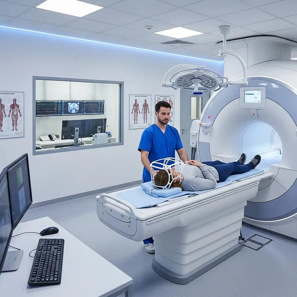
MRI is a powerful imaging tool that uses magnetic fields and radio waves to produce detailed images of the body’s internal structures. It is particularly advantageous for evaluating soft tissue abnormalities and is often used when other imaging modalities are inconclusive. MRI is non-invasive and does not involve ionizing radiation, making it a safer option for certain patients.
In Which Clinical Situations Is MRI Abdomen Preferred?
MRI is preferred in clinical situations where detailed images of soft tissues are required, such as in the evaluation of liver lesions, pancreatic disorders, and certain gynecological conditions. It is also beneficial for assessing complex abdominal masses that may not be clearly defined on CT scans.
What Are the Advantages of MRI Compared to Other Imaging Modalities?
The advantages of MRI over other imaging modalities include its superior soft tissue contrast and the absence of radiation exposure. MRI can provide detailed information about the composition of tissues, which is crucial for accurate diagnosis and treatment planning. Additionally, MRI is particularly useful in monitoring the progression of certain diseases over time.
How Do Doctors Decide Which Scan to Order for Abdominal Pain?
Doctors consider various factors when deciding which scan to order for abdominal pain. These factors include the patient’s clinical presentation, the suspected diagnosis, and the urgency of the situation.
What Factors Influence the Choice Between Ultrasound, CT, and MRI?
The choice between ultrasound, CT, and MRI is influenced by several factors:
- Patient Symptoms: The nature and severity of the symptoms can guide the selection of the appropriate imaging modality.
- Medical History: Previous medical conditions and treatments may affect the choice of imaging.
- Physical Examination Findings: The results of the physical examination can provide critical insights into the underlying cause of abdominal pain.
How Do Imaging Protocols and Preparation Affect Scan Selection?
Imaging protocols and preparation play a significant role in the effectiveness of the scans. Proper preparation can enhance the quality of the images obtained, leading to more accurate diagnoses. For instance, patients may be required to fast before certain scans to ensure clear imaging results.
How Can Patients Prepare for Abdominal Imaging Scans and Book Appointments?
Patients can take several steps to prepare for abdominal imaging scans, ensuring a smooth process and accurate results.
- Preparation Requirements for Abdominal Ultrasound and CT Scans: Fasting: Patients may need to fast for several hours before the scan to reduce the presence of food in the stomach.Clothing and Accessories: It is advisable to wear loose-fitting clothing and avoid jewelry that may interfere with the imaging process.
- How to Book an Abdominal Imaging Appointment with X-Ray Docs?
Booking an appointment with X-Ray Docs is straightforward. Patients can contact the facility directly to schedule their imaging services. It is essential to provide relevant medical information and any previous imaging results to ensure a comprehensive evaluation. X-Ray Docs offers a range of imaging services, including ultrasound, CT scans, MRI, and X-rays, catering to various diagnostic needs.
By understanding the different imaging techniques and their applications, patients can engage more effectively in their healthcare decisions and ensure they receive the most appropriate care for their abdominal pain.