

Chest Imaging for Respiratory Conditions: When Do You Need an X-Ray or CT?
Chest imaging plays a crucial role in diagnosing respiratory conditions, helping healthcare professionals determine the underlying causes of symptoms such as cough, chest pain, and difficulty breathing. Understanding when to opt for an X-ray or a CT scan can significantly impact patient outcomes. This article will explore the indications for using these imaging techniques, their benefits and risks, and how to prepare for these procedures. Additionally, we will discuss how to book chest imaging appointments at X-Ray Docs, a leading diagnostic imaging provider in South Africa specializing in accessible chest imaging services.
Indeed, the critical role of imaging in addressing widespread respiratory health challenges cannot be overstated, underscoring the need for early and precise diagnostic methods.
Role of Imaging in Respiratory Diagnosis & Management
Respiratory problems are a significant public health concern, with various conditions posing a threat to individuals’ respiratory systems. These conditions can range from common illnesses such as pneumonia and bronchitis to more serious diseases like lung cancer and COVID-19. For respiratory disorders to be effectively treated and managed, early identification and precise diagnosis are essential. Traditional methods of diagnosing respiratory problems often involve the use of imaging techniques, such as X-ray images.
The Role of Imaging Techniques in the Diagnosis and Management of Respiratory Problems, 2024
Indications for Using X-Rays and CT Scans in Chest Imaging
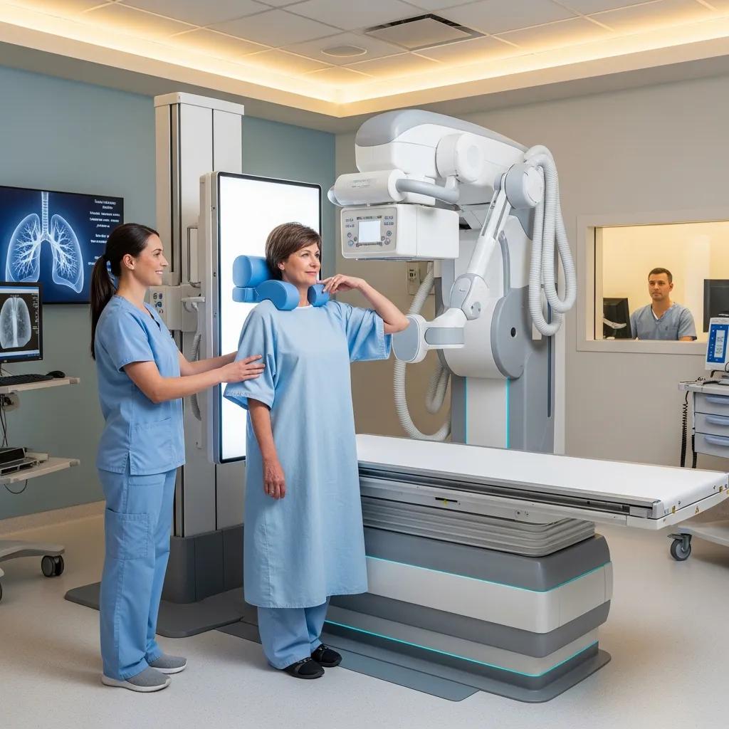
Chest X-rays and CT scans are essential tools in the evaluation of respiratory conditions. They are typically recommended when patients present with specific symptoms or when preoperative assessments are necessary. Common indications for imaging include persistent cough, chest pain, and shortness of breath. These imaging modalities help in monitoring chronic conditions such as COPD and pneumonia, as well as in the evaluation of lung cancer and pulmonary embolism.
Further emphasizing the diagnostic power of imaging, studies show that advanced techniques like ultra-low-dose CT can detect pneumonia even in patients without typical respiratory symptoms.
CXR vs. ULDCT for Pulmonary Infection Diagnosis
Pneumonia can be present in ED patients without respiratory symptoms or signs who have a fever, hypothermia, and/or elevated CRP. ULDCT’s sensitivity is a significant advantage over CXR when pneumonia has to be excluded. Pulmonary imaging in patients with suspected infection but no respiratory symptoms or signs can result in the detection of clinically significant pneumonia. The increased sensitivity of ultra-low-dose chest CT compared to CXR is of added value in vulnerable and immunocompromised patients.
The yield of chest X-ray or ultra-low-dose chest-CT in emergency department patients suspected of pulmonary infection without respiratory symptoms or signs, IAH van den Berk, 2023
X-Ray Docs offers comprehensive chest imaging services, including both X-rays and CT scans, to ensure accurate diagnosis and effective treatment planning.
Chest X-Rays:
Chest X-rays are often the first imaging test performed for respiratory issues. They provide a quick and broad overview of the chest, allowing for the identification of abnormalities such as fluid accumulation, infections, or tumors.
Indications:
Chest X-rays are recommended in various scenarios, including:
- Cough and Chest Pain: Persistent symptoms may indicate underlying conditions that require further investigation.
- Preoperative Assessments: X-rays are often required before surgeries to evaluate lung health.
Benefits:
The advantages of chest X-rays include:
- Quick Procedure: X-rays can be performed in a matter of minutes, providing immediate results.
- Broad Overview of Chest: They help identify a range of conditions, from infections to structural abnormalities.
Potential Risks:
While chest X-rays are generally safe, there are some potential risks to consider:
- Radiation Exposure: Although the dose is low, repeated exposure can accumulate over time.
- Cumulative Risks: Patients should be aware of their overall exposure to radiation from multiple imaging studies.
It is crucial for both practitioners and patients to be aware of the long-term implications of radiation exposure, emphasizing the need for careful risk-benefit assessment in diagnostic imaging.
Radiation Risks in Chest Imaging & Patient Safety
The increasing use and complexity of imaging techniques have not been matched by increasing awareness and knowledge by prescribers and practitioners. Imaging examinations that expose to ionizing radiation provide immense benefits when appropriate, yet they may result in an increased incidence of radiation-induced cancer in the long-term. The radiation issue is relevant not only for the individual patient but also for the community because small individual risks multiplied by millions of examinations become a significant population risk. As recently highlighted by recent European and American Guidelines, the long-term risk associated with radiation exposure should be considered in the risk-benefit assessment behind appropriate prescription of diagnostic testing.
The risk of cumulative radiation exposure in chest imaging and the advantage of bedside ultrasound, L Gargani, 2015
CT Scans:
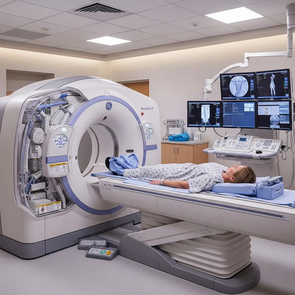
CT scans provide more detailed images of the chest compared to X-rays, making them invaluable for diagnosing complex conditions.
Indications:
CT scans are recommended in specific situations, such as:
- Lung Cancer Diagnosis: They offer detailed images that help in staging and treatment planning.
- Trauma Assessment: CT scans are crucial in evaluating injuries to the chest and lungs.
Benefits:
The benefits of CT scans include:
- Detailed Images: CT scans provide high-resolution images that can reveal subtle abnormalities.
- 3D Reconstructions: They allow for three-dimensional visualization of the chest structures, aiding in precise diagnosis.
Potential Risks:
CT scans also come with risks, including:
- Higher Radiation Exposure: They involve a higher dose of radiation compared to X-rays.
- Cost Considerations: CT scans are generally more expensive than X-rays, which may be a factor for some patients.
What Are the Key Differences Between Chest X-Ray and CT Scan?
Understanding the differences between chest X-rays and CT scans is essential for making informed decisions about imaging.
Key Differences Between Chest X-Ray and CT Scan in Chest Imaging for Respiratory Conditions
Chest X-rays provide a general overview, while CT scans offer detailed cross-sectional images. X-rays are typically used for initial assessments, whereas CT scans are reserved for more complex cases requiring detailed visualization.
How Do Chest X-Rays and CT Scans Differ in Lung Imaging?
The primary difference lies in the level of detail provided. X-rays are effective for identifying obvious issues, while CT scans can detect smaller lesions and provide information about the size and location of abnormalities.
What Are the Benefits and Limitations of Each Imaging Technique?
Both imaging techniques have their unique advantages and limitations.
This comparison highlights the importance of choosing the appropriate imaging technique based on the clinical scenario.
When Should You Get Chest Imaging for Respiratory Diseases?
Chest imaging should be considered when symptoms persist or worsen, particularly in individuals with risk factors for respiratory diseases. Guidelines suggest imaging for patients with chronic cough, unexplained weight loss, or significant changes in respiratory function.
What Are the Indications for Chest X-Ray in Respiratory Conditions?
Chest X-rays are indicated for:
- Evaluation of Symptoms: Persistent cough or chest pain.
- Infection Suspicions: To rule out pneumonia or other infections.
- Chronic Disease Management: Monitoring conditions like COPD.
When Is a Chest CT Scan Recommended for Lung Evaluation?
CT scans are recommended for:
- Trauma Assessment: Evaluating injuries to the chest.
- Cancer Diagnosis: Detailed imaging for staging lung cancer.
- Internal Organ Evaluation: Assessing other thoracic structures.
How Should Patients Prepare for Chest Imaging Procedures?
Preparation for chest imaging is essential to ensure accurate results.
What Steps Are Needed to Prepare for a Chest X-Ray?
Patients should:
- Remove Metal Objects: Jewelry and other items that may interfere with imaging.
- Wear Comfortable Clothing: Loose-fitting clothes are recommended.
How to Prepare for a CT Scan of the Chest?
Preparation for a CT scan includes:
- Dietary Restrictions: Patients may need to avoid food for a few hours before the scan.
- Medication Instructions: Informing the healthcare provider about any medications taken.
How to Book Chest Imaging Appointments at X-Ray Docs in South Africa?
Booking an appointment for chest imaging at X-Ray Docs is straightforward.
What Is the Process for Scheduling a Chest X-Ray or CT Scan?
Patients can schedule appointments by:
- Visiting the Website: Accessing the online booking system.
- Providing Information: Filling out necessary details for the appointment.
What Are the Benefits of Choosing X-Ray Docs for Respiratory Imaging?
Choosing X-Ray Docs offers several advantages:
- Convenient Booking: Easy online scheduling for patients.
- Specialized Imaging: Expertise in chest imaging ensures accurate diagnoses.