Brain CT Scan: What It Detects and What to Expect for Patients
A brain CT scan, or computed tomography scan, is a crucial diagnostic tool that provides detailed images of the brain’s structure. This imaging technique uses X-rays to create cross-sectional views, allowing healthcare professionals to identify various neurological conditions. Patients often seek a brain CT scan to diagnose issues such as strokes, tumors, or traumatic injuries. Understanding what a brain CT scan detects and what to expect during the procedure can alleviate anxiety and prepare patients for their appointments. This article will cover the conditions detectable by brain CT scans, the procedure itself, preparation guidelines, and safety considerations. For more information, you can visit X-Ray Docs.
Further elaborating on the technical aspects and patient advantages, research highlights how CT scanners function and the benefits they offer.
How Brain CT Scans Work & Patient Benefits
CT scanners create cross-sectional images by measuring x-ray attenuation properties of the body from many different directions. With high quality CT imaging being performed more frequently, patients can benefit from a quicker and more accurate diagnosis and precise anatomic information for planning therapeutic procedures.
Strategies for reducing radiation dose in CT, CH McCollough, 2009
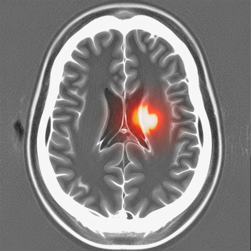
What Conditions Can a Brain CT Scan Detect?
A brain CT scan is instrumental in diagnosing several critical conditions. It can effectively identify:
- Stroke: CT scans can quickly reveal the presence of a stroke, allowing for timely intervention.
- Brain Tumors: These scans help in detecting tumors, assessing their size, and determining their location.
- Hemorrhages: A brain CT scan can identify bleeding within the brain, which is crucial for emergency treatment.
The ability to diagnose these conditions promptly is vital, as it can significantly impact treatment outcomes and patient recovery.
Specialized CT techniques, such as perfusion CT, provide even deeper insights into conditions like strokes and tumors, as detailed by experts.
Brain Perfusion CT: Diagnosing Strokes & Tumors
By providing quantitative measurements of cerebral blood flow (CBF) and cerebral blood volume (CBV), dynamic perfusion computed tomography (p-CT) allows visualisation of cerebral autoregulation mechanisms and represents a fast, available and reliable imaging option for assessing cerebral perfusion. Thanks to its feasibility in emergency settings, p-CT is considered most useful, in combination with CT angiography, in acute ischaemic patients, as it is able to provide a fast and noninvasive assessment of cerebral perfusion impairment. In addition, p-CT can play a diagnostic role in other types of cerebrovascular disease to assess functional reserve, and in intracranial neoplasms, where it has a role in diagnosis, grading, biopsy guidance, and follow-up during treatment.
Brain perfusion CT: principles, technique and clinical applications, M Wintermark, 2007
How Does a Brain CT Scan Identify Stroke and Hemorrhage?
Brain CT scans utilize advanced imaging technology to detect strokes and hemorrhages. The process involves taking multiple X-ray images from different angles, which are then processed to create detailed cross-sectional images of the brain.
When a stroke occurs, either due to a blockage (ischemic stroke) or bleeding (hemorrhagic stroke), the CT scan can show changes in brain tissue and blood flow. Rapid imaging is essential, as it allows healthcare providers to determine the type of stroke and initiate appropriate treatment quickly. You can find more information about brain CT scans on specialized radiology websites.
Can Brain Tumors and Traumatic Injuries Be Detected by CT Imaging?
Yes, brain tumors and traumatic injuries can be effectively detected through CT imaging. The high-resolution images produced by a CT scan allow for the visualization of abnormal growths and structural changes in the brain.
CT scans are particularly useful in emergency situations where traumatic brain injuries are suspected. They can reveal fractures, contusions, and other injuries that may not be visible through other imaging techniques. This capability makes CT scans a preferred choice in acute care settings.
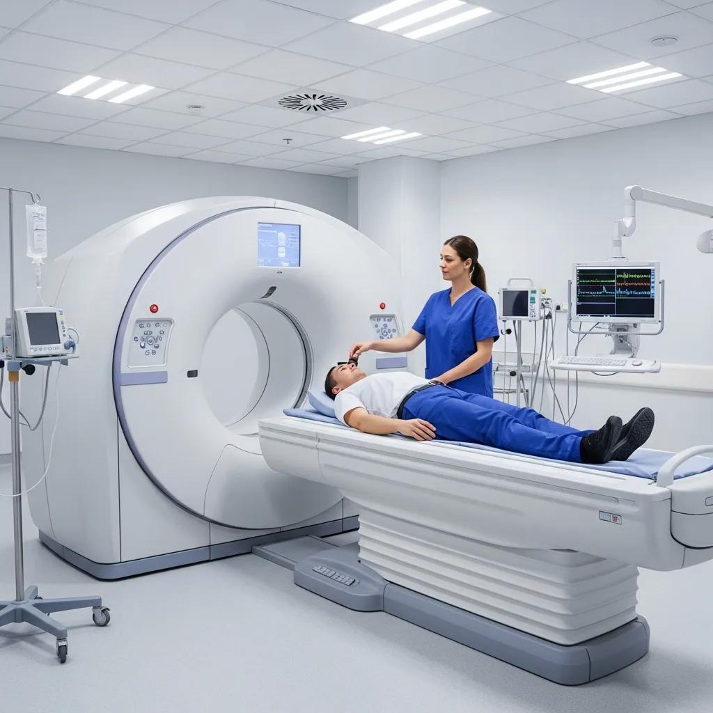
What to Expect During the Head CT Scan Procedure
Understanding what to expect during a head CT scan can help ease patient anxiety. The procedure is typically quick and straightforward.
Proper patient positioning is crucial for obtaining clear and accurate images during the scan, as outlined in clinical guidelines.
Brain CT Scan: Patient Positioning Guide
Patients should be asked to lie supine on the CT scan table with their heart located in the center of the gantry (slightly lateral to midline). Table height should be adjusted until the
Patient preparation and scanning techniques, S Abbara, 2010
Patients will be asked to lie on a table that slides into the CT scanner. The machine will rotate around the head, capturing images from various angles. It is essential to remain still during the scan to ensure clear images. The entire process usually takes about 5 to 30 minutes, depending on the specific requirements of the scan.
How Is the Brain CT Scan Performed Step-by-Step?
The brain CT scan procedure involves several key steps:
- Preparation: Patients may be asked to remove any metal objects and change into a hospital gown.
- Positioning: The patient lies on a table, and a strap may be used to keep the head still.
- Imaging: The CT scanner rotates around the head, taking multiple images.
- Post-Scan: After the scan, patients can typically resume normal activities unless otherwise instructed.
What Are the Typical Duration and Patient Experience?
The duration of a brain CT scan is generally brief, lasting between 5 to 30 minutes. Patients often report that the experience is relatively comfortable, with minimal discomfort.
During the scan, patients may hear a series of clicking noises as the machine operates. It is important to follow the technician’s instructions, especially regarding breath-holding, to ensure the best possible images are obtained.
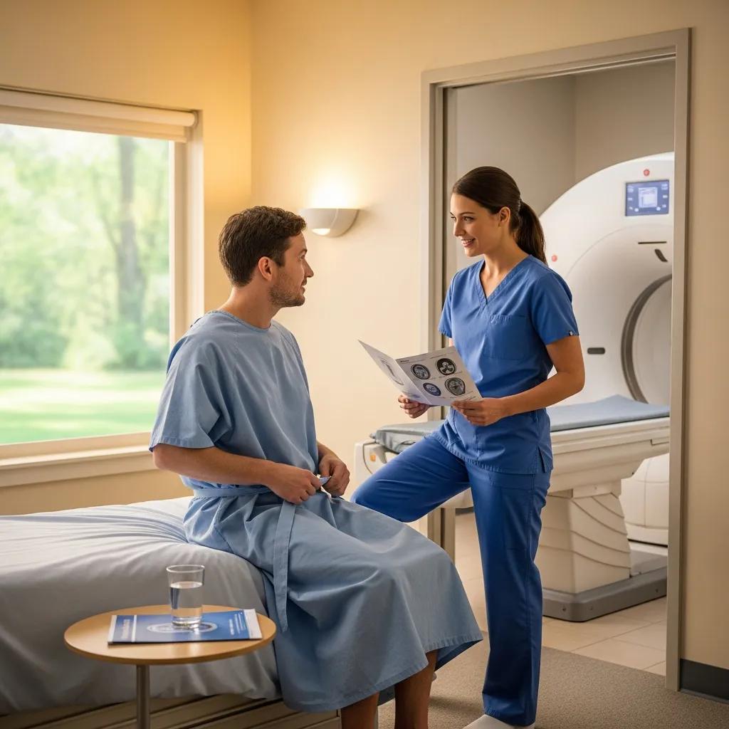
How to Prepare for a Brain CT Scan Appointment
Preparation for a brain CT scan is essential to ensure accurate results. Patients should follow specific guidelines to facilitate the process.
What Are the Preparation Guidelines for a Head CT Scan?
Patients are typically advised to:
- Avoid Food and Drink: Fasting for a few hours before the scan may be required, especially if contrast material is used.
- Inform the Technician: Notify the technician of any allergies, particularly to iodine or contrast materials.
- Wear Comfortable Clothing: Loose-fitting clothes without metal fasteners are recommended.
Are There Specific Instructions Regarding Medications or Food?
Patients should consult their healthcare provider regarding any medications they are taking. Some medications may need to be paused before the scan, particularly those affecting blood clotting.
Additionally, patients should follow any dietary restrictions provided by their healthcare team to ensure optimal imaging results.
What Are the Risks and Safety Considerations of Brain CT Scans?
While brain CT scans are generally safe, it is essential to be aware of potential risks and safety considerations.
What Are the Potential Radiation Risks Associated with Brain CT?
CT scans involve exposure to ionizing radiation, which can pose risks, particularly with repeated exposure. However, the benefits of accurate diagnosis often outweigh these risks.
Healthcare providers take precautions to minimize radiation exposure, such as using the lowest effective dose and limiting the number of scans performed. For more detailed information on safety protocols, visit X-Ray Docs.
How Does X-Ray Docs Ensure Patient Safety During Imaging?
X-Ray Docs prioritizes patient safety by adhering to strict protocols and guidelines. The facility employs certified technicians and uses state-of-the-art equipment to ensure high-quality imaging while minimizing risks.
Regular maintenance and calibration of the CT machines further enhance safety, ensuring that patients receive the best possible care during their imaging procedures.