CT Scan for Chest Pain Diagnosis: When and Why You Need Imaging
Chest pain can be a distressing symptom, often leading to urgent medical evaluations. Understanding when a CT scan is necessary for diagnosing chest pain is crucial for timely and effective treatment. This article will explore the indications for a CT scan, its benefits, potential risks, and the specific symptoms that warrant imaging. Additionally, we will discuss the different imaging tests available for chest pain and how to prepare for a chest CT scan. By the end, you will have a comprehensive understanding of when to seek imaging services for chest pain and how “X-Ray Docs” can assist you in this process.
Indications for a CT Scan
A CT scan is often indicated in specific scenarios where chest pain may suggest serious underlying conditions. These include:
- Pulmonary Embolism Detection: A CT scan can quickly identify blood clots in the lungs, which can be life-threatening.
- Cancer Diagnosis: Imaging is essential for detecting tumors in the lungs or surrounding tissues.
- Trauma Assessment: In cases of chest trauma, a CT scan helps evaluate internal injuries.
Recognizing these indications can lead to prompt diagnosis and treatment, potentially saving lives.
Further guidance on diagnosing the cause of chest pain suggests that specific risk assessments and blood tests can help determine the necessity of a helical CT for pulmonary embolism.
When Helical CT is Needed for Pulmonary Embolism Diagnosis
Risk of pulmonary embolism can be determined with a simple prediction rule, and a d-dimer assay can help determine whether further evaluation with helical computed tomography or venous ultrasound is needed.
Diagnosing the cause of chest pain, 2005
Benefits of a CT Scan
CT scans offer several advantages in the evaluation of chest pain:
- Detailed Imaging: CT scans provide high-resolution images that allow for a comprehensive view of the chest structures.
- Rapid Diagnosis: The speed of CT imaging can facilitate quick decision-making in emergency situations.
- 3D Reconstructions: Advanced imaging techniques enable 3D reconstructions, aiding in the assessment of complex anatomical structures.
These benefits make CT scans a valuable tool in the diagnostic process for chest pain.
Potential Risks
While CT scans are generally safe, there are potential risks to consider:
- Radiation Exposure: CT scans involve exposure to ionizing radiation, which can increase cancer risk over time.
- Contrast Material Reactions: Some patients may experience allergic reactions to the contrast material used in CT scans.
- Pregnancy Considerations: Pregnant women should avoid unnecessary radiation exposure, making alternative imaging methods preferable when possible.
Understanding these risks is essential for informed decision-making regarding imaging.
When Should You Get a Chest CT Scan for Chest Pain?
Certain symptoms indicate the need for a CT scan when experiencing chest pain. These include:
- Chest Pain Severity: Severe or worsening chest pain should prompt immediate evaluation.
- Duration of Symptoms: Persistent chest pain lasting more than a few minutes warrants further investigation.
- Associated Symptoms: Symptoms such as shortness of breath, dizziness, or sweating alongside chest pain may indicate a serious condition.
Recognizing these symptoms can help patients seek timely medical attention.
What Chest Pain Symptoms Indicate the Need for a CT Scan?

Specific types of chest pain can signal the necessity for a CT scan. For instance, sharp, stabbing pain that radiates to the arm or jaw may suggest a heart attack. Additionally, pain accompanied by difficulty breathing or a feeling of impending doom should prompt immediate medical evaluation. Understanding these indicators can lead to quicker diagnoses and better outcomes.
Research further emphasizes the critical role of cardiac CT in evaluating acute chest pain and diagnosing acute coronary syndrome.
Cardiac CT for Acute Chest Pain & Acute Coronary Syndrome Diagnosis
As the use of CT increases in the imaging of patients with acute chest pain, it is important to understand its role in the diagnosis of an acute coronary syndrome. If it is determined that particular multidetector CT findings are highly predictive of an acute coronary syndrome, then the patient can be treated appropriately.
Cardiac CT in emergency department patients with acute chest pain, U Hoffmann, 2006
How Does CT Angiography Help Diagnose Pulmonary Embolism and Aortic Dissection?
CT angiography is a specialized imaging technique that enhances the visualization of blood vessels. It is particularly useful in diagnosing conditions like pulmonary embolism and aortic dissection. By injecting contrast material, CT angiography allows for detailed images of the blood vessels, helping to identify blockages or tears. This rapid and accurate assessment is crucial in emergency settings.
Indeed, the advancements in CT angiography have solidified its position as the preferred diagnostic tool for suspected acute pulmonary embolism.
CT Angiography: Study of Choice for Suspected Pulmonary Embolism
The improved spatial resolution, speed of diagnosis, reassurance that negative CT angiograms and venograms can be relied upon, and the ability to detect alternative diagnoses has made CT angiography or CT angiography with CT venography the study of choice for suspected acute pulmonary embolism.
Management of Suspected Acute Pulmonary Embolism in the Era of CT Angiography: A Statement from the Fleischner Society1, 2007
What Are the Different Imaging Tests for Chest Pain?
Several imaging tests are available for evaluating chest pain, each with its unique advantages:
- CT Scans: Provide detailed cross-sectional images of the chest.
- Chest X-rays: Useful for initial assessments but less detailed than CT scans.
- MRI for Chest Pain: Offers good soft tissue contrast but is less commonly used for acute chest pain evaluations.
Understanding the differences between these modalities can help in selecting the appropriate imaging test based on the clinical scenario.
How Do CT Scans Compare to X-rays and MRI in Chest Pain Diagnosis?
CT scans are often preferred over X-rays and MRIs for chest pain diagnosis due to their speed and detail. While X-rays can identify some abnormalities, they lack the resolution needed for a comprehensive assessment. MRIs, although excellent for soft tissue evaluation, are not typically used in acute settings due to longer scan times. Therefore, CT scans are often the go-to choice for rapid and accurate diagnosis.
When Is Non-Invasive Imaging Preferred Over Other Modalities?
Non-invasive imaging techniques, such as CT scans, are preferred in many scenarios due to their safety and effectiveness. They minimize patient discomfort and avoid the risks associated with invasive procedures. Additionally, non-invasive imaging can provide critical information quickly, which is essential in emergency situations. This approach prioritizes patient safety while ensuring accurate diagnoses.
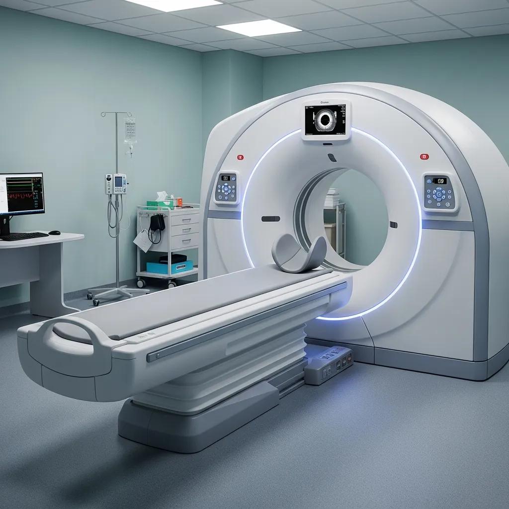
How Is a Chest CT Scan Performed and What Should Patients Expect?
A chest CT scan is a straightforward procedure that typically takes about 10-30 minutes. Patients lie on a table that slides into the CT scanner, which takes multiple images of the chest from different angles.
What Is the Preparation Process for a Chest CT Scan?

Preparation for a chest CT scan may include:
- Dietary Restrictions: Patients may be advised to avoid food or drink for a few hours before the scan.
- Clothing Considerations: Loose-fitting clothing without metal fasteners is recommended.
- Medication Instructions: Patients should inform their doctor about any medications they are taking, especially if they have allergies to contrast material.
Following these guidelines can help ensure a smooth imaging process.
What Are the Risks and Benefits Associated with Chest CT Imaging?
The decision to undergo a chest CT scan involves weighing the risks and benefits.
| Risk | Description | Impact Level |
|---|---|---|
| Radiation Exposure | Potential increase in cancer risk | Moderate |
| Contrast Reactions | Allergic reactions to contrast material | Low |
| Pregnancy Risks | Avoidance of radiation exposure | High |
Understanding these factors can help patients make informed choices about their imaging options.
How to Book Professional Chest Pain Imaging Services at X-Ray Docs
Booking a CT scan for chest pain at X-Ray Docs is a straightforward process. Patients can expect to provide necessary documentation and insurance information during scheduling.
What Are the Steps to Schedule a CT Scan for Chest Pain Diagnosis?
To schedule a CT scan for chest pain diagnosis, follow these steps:
- Referral Requirements: Obtain a referral from a healthcare provider.
- Booking Process: Contact X-Ray Docs to schedule an appointment.
- What to Expect During the Visit: Arrive on time, bring necessary documents, and follow pre-scan instructions.
This streamlined process ensures that patients receive timely and efficient care.
How Does X-Ray Docs Ensure Patient-Centered Care and Expertise?
X-Ray Docs prioritizes patient-centered care by employing professional staff and maintaining a streamlined process. The focus is on patient comfort and ensuring that each individual receives the highest quality of care during their imaging experience. This commitment to excellence sets X-Ray Docs apart in the field of diagnostic imaging.