Dental X-Rays vs Medical X-Rays: Key Differences and Patient Guide
X-rays are a vital tool in modern medicine, providing crucial insights into both dental and medical health. Understanding the differences between dental X-rays and medical X-rays can help patients make informed decisions about their healthcare. This article will explore the fundamental principles of X-ray technology, the specific applications of both types of X-rays, and the key differences that set them apart. Many patients may feel anxious about radiation exposure, but knowing how these imaging techniques work can alleviate concerns. We will also discuss the safety protocols in place to protect patients during these procedures. Finally, we will highlight why choosing a trusted provider like Tesla Radiological Services is essential for your imaging needs.
Indeed, the foundational role of diagnostic radiography in both medical and dental fields is paramount for accurate diagnosis and effective treatment planning.
Diagnostic Radiography in Medical & Dental Practice
The rapid progress of medical and dental science with the invention of various drugs have benefited the mankind. The proper and correct diagnosis of diseases is the primary necessity before the treatment. The different imaging modality plays an important role in clinical diagnosis, teaching and in the field of research. With advent of modern technology in imaging medical imaging has also greatly influenced. A diagnostic radiograph has become a hotspot in diagnosis and the clinical applications. The technology advancement has greatly influenced the medical imaging in many ways even in the aspect of storage of images. Oral and maxillofacial radiography is the art of recording images of a patient’s oral and associated structures. Radiographic examinations play an essential part of dental practice.
The road to radiation safety and ALARA: A review, BB Joseph, 2021
What Are X-Rays and How Do They Work?
X-rays are a form of electromagnetic radiation that can penetrate various materials, including human tissue. This property allows them to create images of the internal structures of the body, which are essential for diagnosis and treatment planning. The basic science behind X-ray imaging involves the use of an X-ray machine that emits radiation, which passes through the body and is captured on a film or digital sensor. The resulting images reveal the density of tissues, with denser materials like bones appearing white and softer tissues appearing darker.
What is the basic science behind X-ray imaging?
The fundamental principle of X-ray imaging lies in the interaction between X-rays and matter. When X-rays pass through the body, they are absorbed by different tissues at varying rates. Dense tissues, such as bones, absorb more X-rays and appear white on the image, while softer tissues allow more X-rays to pass through, resulting in darker areas. This contrast is what enables healthcare professionals to identify abnormalities, fractures, or diseases.
How do dental and medical X-rays utilize radiation for diagnosis?
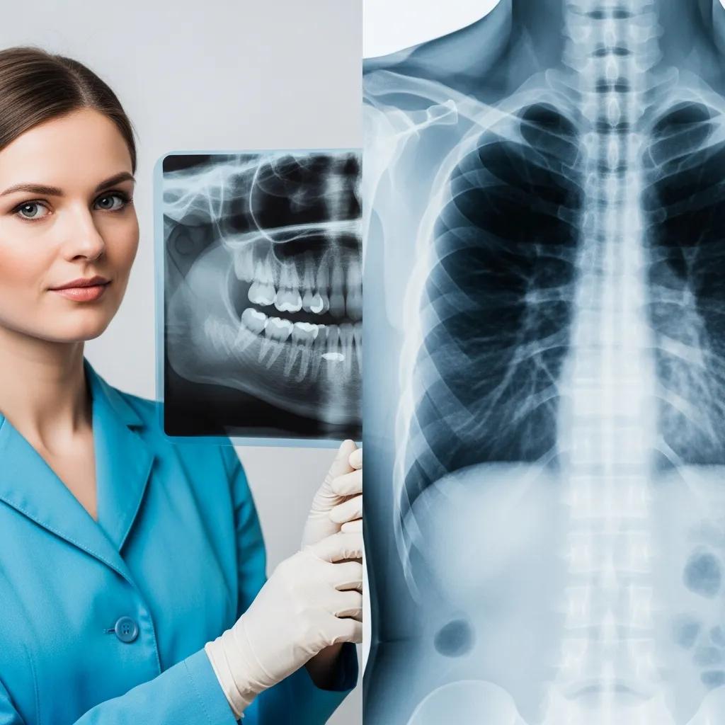
Both dental and medical X-rays utilize radiation to create images, but they differ in their applications and the areas they target. Dental X-rays focus on the teeth, gums, and jawbone, while medical X-rays can examine various body parts, including the chest, abdomen, and limbs. The radiation doses used in dental X-rays are typically much lower than those in medical X-rays, reflecting the smaller target area and the need for precise imaging in dental care.
What Are Medical X-Rays and Their Common Applications?
Medical X-rays are a crucial diagnostic tool used to visualize the internal structures of the body. They are commonly employed to diagnose a wide range of conditions, including fractures, infections, and tumors. Medical X-rays can be used to assess various body parts, such as the chest for lung conditions, the abdomen for digestive issues, and the limbs for bone injuries.
Which body parts and conditions do medical X-rays diagnose?
- Fractures: Identifying broken bones in various parts of the body.
- Infections: Detecting pneumonia or other infections in the lungs.
- Tumors: Visualizing abnormal growths in different organs.
How does Tesla Radiological Services provide advanced medical imaging?
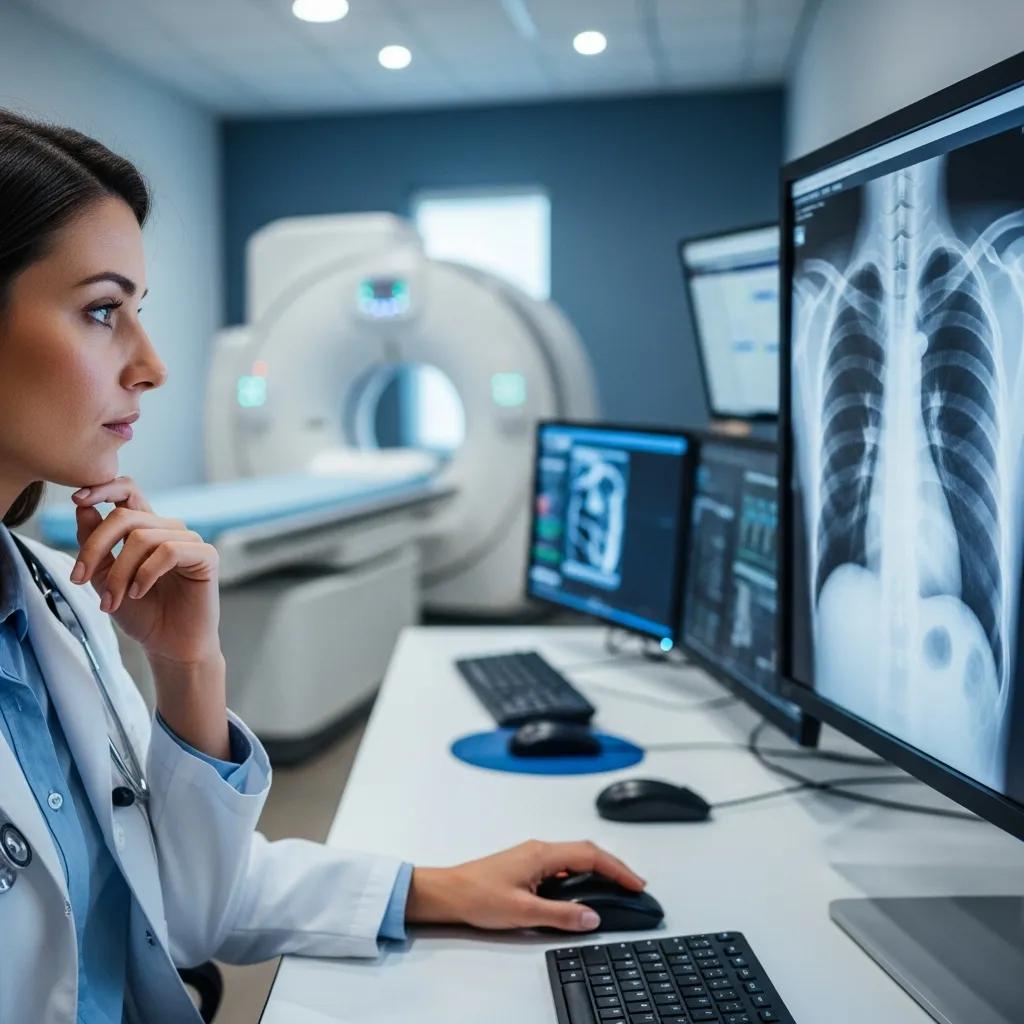
Tesla Radiological Services offers a comprehensive range of medical imaging services, including X-rays, CT scans, and MRIs. The facility is equipped with state-of-the-art technology and staffed by experienced professionals who prioritize patient care. By utilizing advanced imaging techniques, Tesla ensures accurate diagnoses and effective treatment planning for various medical conditions.
What Are Dental X-Rays and Their Specific Uses?
Dental X-rays are specialized imaging techniques used to examine the teeth, gums, and surrounding structures. They play a vital role in diagnosing oral health issues, planning treatments, and monitoring the progress of dental procedures. Different types of dental X-rays are employed based on the specific needs of the patient.
What types of dental X-rays are commonly used?
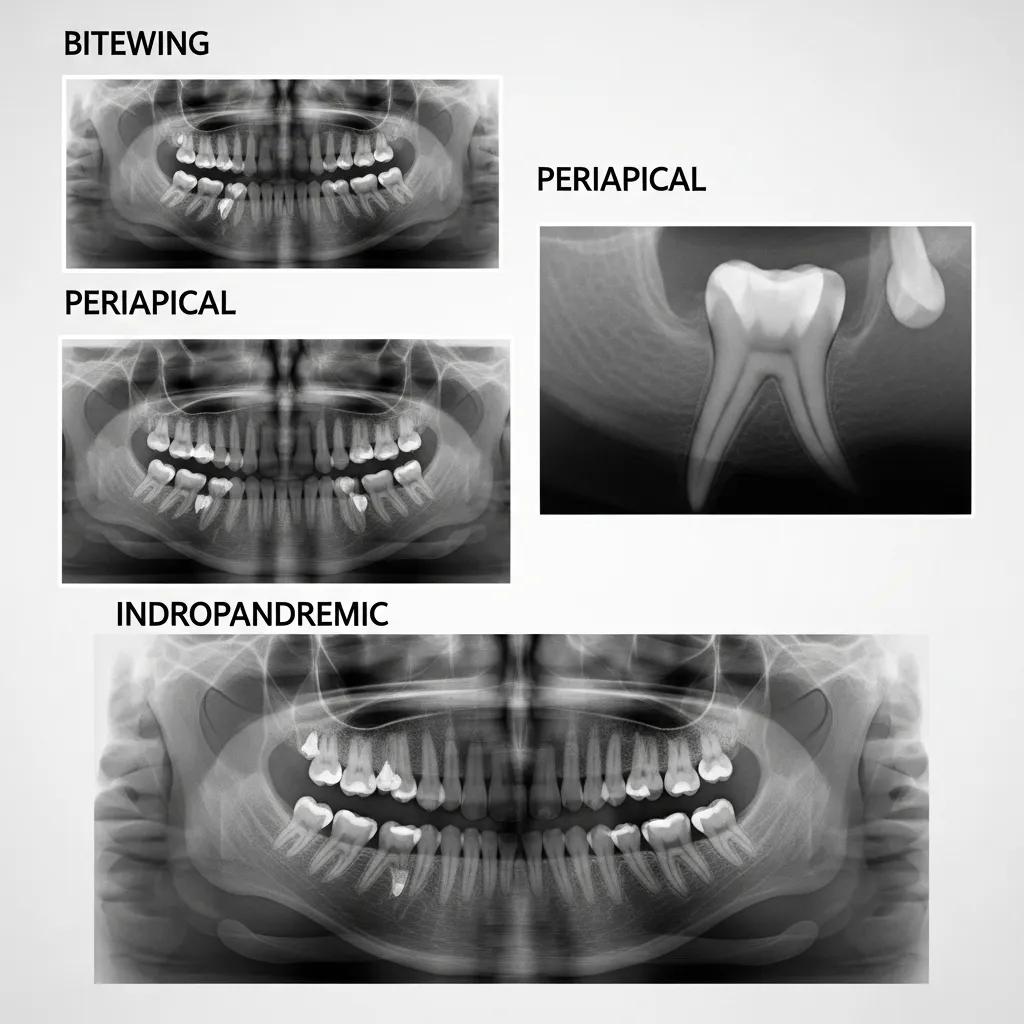
- Bitewing X-rays: These capture the upper and lower teeth in one area of the mouth, helping to detect cavities between teeth.
- Periapical X-rays: These focus on one or two specific teeth, providing detailed images of the tooth’s root and surrounding bone.
- Panoramic X-rays: These offer a broad view of the entire mouth, including all teeth, jaws, and surrounding structures.
How do dental X-rays detect oral health issues?
- Cavities: Early detection of decay that may not be visible during a routine examination.
- Bone loss: Assessing the health of the jawbone and surrounding structures.
- Gum disease: Identifying signs of periodontal disease that may require treatment.
What Are the Key Differences Between Dental and Medical X-Rays?
While both dental and medical X-rays utilize similar technology, they differ significantly in their target areas, equipment, and radiation doses. Understanding these differences can help patients appreciate the specific applications of each type of X-ray.
How do target areas and equipment differ between dental and medical X-rays?
The target areas for dental and medical X-rays are distinct. Dental X-rays focus on the oral cavity, while medical X-rays can examine various body parts. The equipment used also varies; dental X-ray machines are typically smaller and designed for precise imaging of teeth and gums, whereas medical X-ray machines are larger and capable of producing images of the entire body.
What are the differences in radiation dose and diagnostic focus?
Dental X-rays generally involve much lower radiation doses compared to medical X-rays. This is due to the smaller area being imaged and the need for precision in dental diagnostics. Medical X-rays, on the other hand, may require higher doses to capture detailed images of larger body parts. Understanding these differences is crucial for patients concerned about radiation exposure.
How Safe Are Dental and Medical X-Rays Regarding Radiation Exposure?
Safety is a primary concern for patients undergoing X-ray procedures. Both dental and medical X-rays are designed with safety protocols to minimize radiation exposure while ensuring accurate imaging.
The increasing use of medical imaging underscores the critical importance of patient safety and understanding the associated risks of ionizing radiation.
Patient Safety & Ionizing Radiation Risk in Medical Imaging
Medical imaging is a valuable clinical tool which has led to its growing utilization. Errors in radiology may occur anywhere along the course of a study being ordered, through its acquisition, interpretation, and communication. One specific area of focus for safety in radiology is exposure to ionizing radiation, which has associated immediate and long-term exposure risk.
Patient safety in radiology and medical imaging, A Ding, 2023
What radiation safety protocols minimize patient risk?
- ALARA Principle: Keeping radiation exposure “As Low As Reasonably Achievable” to minimize risk.
- Lead aprons and thyroid collars: Using protective gear to shield sensitive areas of the body from unnecessary radiation.
- Digital technology: Employing digital X-ray systems that require less radiation than traditional film methods.
How do digital X-rays reduce radiation compared to traditional methods?
Digital X-rays significantly reduce radiation exposure compared to traditional film X-rays. The advanced technology used in digital imaging allows for higher sensitivity, meaning less radiation is needed to produce a clear image. This innovation not only enhances patient safety but also improves the overall quality of diagnostic imaging.
Beyond technological advancements, ongoing efforts are crucial to address public concerns and educate both patients and practitioners on minimizing radiation exposure from medical X-rays.
Reducing Medical X-Ray Radiation & Public Awareness
With the increasing frequency of X‑ray examinations in clinical medicine, public concern regarding the harm caused by exposure to X‑ray radiation is also increasing. However, some physicians are not completely aware of the dangers of exposure to X‑ray irradiation. Individuals specialized in this field, including physicians, have a better understanding of these dangers, which limits the use of X‑rays in medicine. The present study aimed to address strategies for reducing the harm caused by exposure to medical X‑rays and increase public awareness regarding X‑ray radiation.
Recommendations for reducing exposure to medical X-ray irradiation, 2022
Why Choose Tesla Radiological Services for Your Imaging Needs?
When it comes to medical imaging, choosing a trusted provider is essential for ensuring accurate diagnoses and effective treatment. Tesla Radiological Services stands out as a comprehensive medical imaging provider in South Africa, offering a wide array of radiological services.
What advanced imaging technologies and expertise does Tesla offer?
Tesla Radiological Services utilizes state-of-the-art imaging technologies, including digital X-rays, CT scans, and MRIs. The facility is staffed by experienced radiologists and technicians who are dedicated to providing high-quality care. Their expertise ensures that patients receive accurate diagnoses and personalized treatment plans tailored to their needs.
How can patients conveniently book appointments for X-ray services?
Patients can easily book appointments for X-ray services at Tesla Radiological Services by contacting the facility directly. The staff is available to assist with scheduling and answer any questions regarding the imaging process. This commitment to patient care ensures a seamless experience from start to finish.
| Type of X-Ray | Target Area | Typical Radiation Dose |
|---|---|---|
| Dental X-ray | Teeth and gums | Very low (0.005 mSv) |
| Medical X-ray | Various body parts | Low to moderate (0.1-10 mSv) |