What Is a Chest X-Ray and How to Interpret Its Findings?
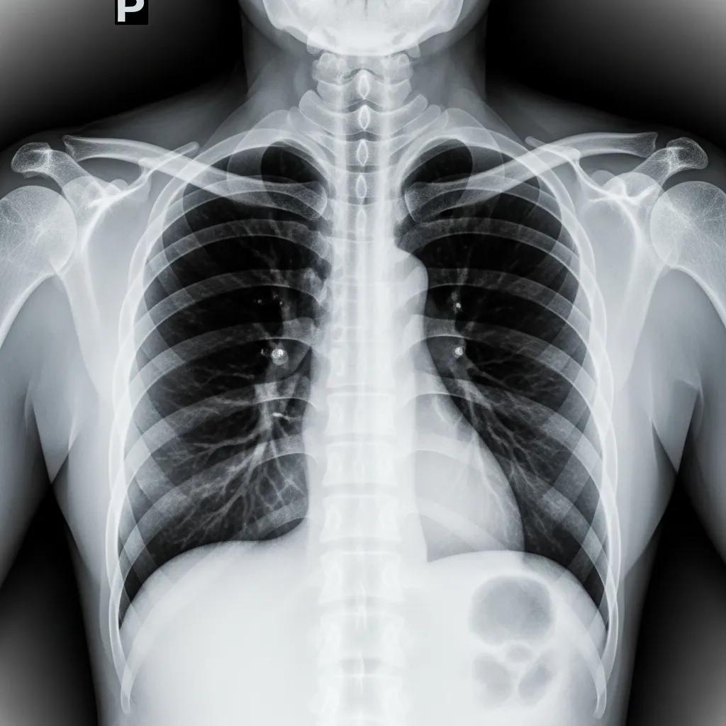
A chest X-ray is a vital diagnostic tool used in medical imaging to visualize the structures within the thoracic cavity, including the lungs, heart, and surrounding tissues. This non-invasive procedure employs electromagnetic radiation to create images that help healthcare professionals assess various conditions. Understanding the interpretation of chest X-rays is crucial for accurate diagnosis and treatment planning. In this article, we will explore the procedure of chest X-rays, what they reveal about lung and heart conditions, and how to prepare for and interpret the findings effectively.
What Is a Chest X-Ray and How Is the Procedure Performed?
A chest X-ray is a type of radiographic imaging that captures detailed images of the chest area. The procedure involves the patient standing or sitting in front of an X-ray machine, where a small amount of radiation is passed through the body to create images on a film or digital detector. The primary purpose of a chest X-ray is to identify abnormalities in the lungs, heart, and other structures within the thorax.
During the procedure, the patient may be asked to take a deep breath and hold it for a few seconds to ensure clear images. The entire process typically takes only a few minutes, making it a quick and efficient diagnostic tool.
Chest X-Ray Findings: Normal Anatomy vs Common Abnormalities
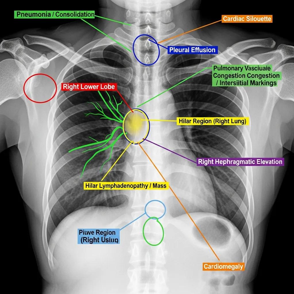
Chest X-rays can reveal a variety of findings, ranging from normal anatomical structures to significant abnormalities. Common findings include:
Advancements in medical imaging are also exploring automated methods to identify conditions like cardiomegaly from chest X-ray images.
Automatic Cardiomegaly Detection on Chest X-Rays
used for the automatic detection of cardiomegaly on chest X-ray images. Results of cardiomegaly despite the normal size of the heart silhouette.
Identifying cardiomegaly in chest X-rays: a cross-sectional study of evaluation and comparison between different transfer learning methods, C Malamateniou, 2021
- Lung Infections: Pneumonia or other infections may appear as areas of increased opacity in the lung fields.
- Fluid Accumulation: Conditions like pleural effusion can be identified by the presence of fluid in the pleural space.
- Tumors: Abnormal masses or nodules may indicate the presence of lung cancer or other malignancies.
- Heart Enlargement: Cardiomegaly can be assessed by examining the size and shape of the heart silhouette.
Understanding these findings is essential for healthcare providers to determine the appropriate course of action for patients.
Indeed, the chest X-ray is widely recognized as a primary method for diagnosing cardiomegaly, or an enlarged heart.
Chest X-Ray for Cardiomegaly Diagnosis
a chest x-ray is the most common way to test for cardiomegaly. In spite of this, the cardiac silhouette is enlarged with a left.
Design and implementation of diagnosis system for cardiomegaly from clinical chest X-ray reports, OA Ogungbe, 2022
| Finding | Description | Clinical Significance |
|---|---|---|
| Lung Infections | Areas of increased opacity | Indicates possible pneumonia or infection |
| Fluid Accumulation | Presence of fluid in pleural space | Suggests pleural effusion or other conditions |
| Tumors | Abnormal masses or nodules | May indicate malignancy or other serious conditions |
| Heart Enlargement | Enlarged heart silhouette | Suggests heart disease or other cardiovascular issues |
These findings highlight the importance of chest X-rays in diagnosing and managing various thoracic conditions.
However, the precise diagnostic accuracy of measurements like the cardiothoracic ratio for detecting specific conditions such as left ventricular enlargement continues to be a subject of ongoing research.
Cardiothoracic Ratio Accuracy in LV Enlargement Detection
The diagnostic accuracy of the cardiothoracic ratio on chest X-ray to detect left ventricular (LV) enlargement has not been well defined despite its traditional association with cardiomegaly. We aimed to determine whether the cardiothoracic ratio can accurately predict LV enlargement based on indexed linear measurements of the LV on transthoracic echocardiography (TTE).
Relationship between enlarged cardiac silhouette on chest X-ray and left ventricular size on transthoracic echocardiography, J Yim, 2022
Which Lung and Heart Conditions Can a Chest X-Ray Detect?
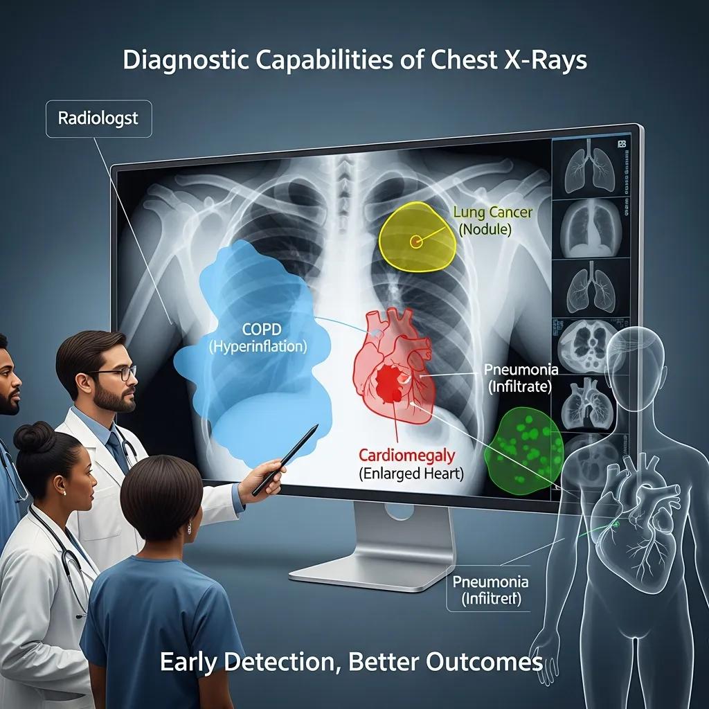
Chest X-rays are instrumental in detecting a range of lung and heart conditions. Some of the key conditions include:
- Chronic Obstructive Pulmonary Disease (COPD): X-rays can show hyperinflation of the lungs and other changes associated with COPD.
- Congestive Heart Failure: Signs of fluid overload and heart enlargement can be visualized.
- Lung Cancer: Early detection of tumors can significantly impact treatment outcomes.
- Tuberculosis: Characteristic patterns of lung involvement can be identified.
By recognizing these conditions through chest X-rays, healthcare providers can initiate timely interventions.
How to Interpret Chest Radiograph Abnormalities?
Interpreting chest X-ray abnormalities requires a systematic approach. Radiologists typically follow these steps:
- Assess the Quality of the X-ray: Ensure the image is clear and properly exposed.
- Evaluate the Anatomy: Identify normal structures and their positions.
- Look for Abnormalities: Note any deviations from normal anatomy, such as masses or fluid levels.
- Consider Clinical Context: Correlate findings with the patient’s symptoms and medical history.
This structured approach aids in accurate diagnosis and effective communication of findings to the healthcare team.
When Is a Chest X-Ray Recommended? Indications and Uses
Chest X-rays are commonly recommended for various clinical indications, including:
- Evaluation of Symptoms: Patients presenting with cough, chest pain, or shortness of breath may require a chest X-ray to identify underlying causes.
- Preoperative Assessment: Surgeons often request chest X-rays before procedures to evaluate lung health.
- Monitoring Disease Progression: Patients with known lung or heart conditions may undergo regular X-rays to monitor changes over time.
These indications underscore the versatility of chest X-rays in clinical practice.
What Are the Common Medical Reasons for Ordering a Chest X-Ray?
Several medical reasons prompt healthcare providers to order chest X-rays, including:
- Infection Suspicions: To confirm or rule out pneumonia or other respiratory infections.
- Trauma Assessment: Following chest injuries, X-rays help identify fractures or internal injuries.
- Chronic Disease Management: Monitoring conditions like COPD or heart failure requires periodic imaging.
These reasons highlight the critical role of chest X-rays in patient care.
How Does a Chest X-Ray Aid in Diagnosing Thoracic Conditions?
Chest X-rays provide essential information for diagnosing thoracic conditions by offering a visual representation of the chest’s internal structures. They help in:
- Identifying Pathologies: X-rays can reveal various diseases affecting the lungs and heart.
- Guiding Further Testing: Abnormal findings may necessitate additional imaging or diagnostic procedures.
- Assessing Treatment Efficacy: Follow-up X-rays can evaluate the effectiveness of treatments for lung or heart conditions.
This diagnostic capability is invaluable in managing patient health.
How to Book a Chest X-Ray at X-Ray Docs and What to Expect
Booking a chest X-ray at X-Ray Docs is a straightforward process designed to facilitate patient access to diagnostic imaging services. Patients can schedule an appointment through the X-Ray Docs website, where they will find options for various radiographic services, including chest X-rays.
During the visit, patients can expect a professional environment where trained radiologists will conduct the procedure efficiently. The staff will guide patients through the process, ensuring comfort and clarity regarding what to expect.
For those seeking a reliable imaging service, X-Ray Docs specializes in diagnostic radiology, including chest X-rays, and is committed to providing high-quality care.
What Should Patients Expect During Their Visit to X-Ray Docs?
During a visit to X-Ray Docs for a chest X-ray, patients can anticipate the following:
- Professional Staff: Trained radiologists will perform the X-ray, ensuring a smooth experience.
- Quick Procedure: The X-ray process typically lasts only a few minutes.
- Post-Procedure Guidance: Patients will receive information on when to expect results and any necessary follow-up.
This streamlined approach ensures that patients receive timely and effective diagnostic services.
To learn more about chest X-rays and other diagnostic services, visit X-Ray Docs today.