What Is Fluoroscopy? Uses, Procedure, and Safety Explained
Fluoroscopy is a medical imaging technique that provides real-time visualization of internal structures and functions within the body. By utilizing X-ray technology, fluoroscopy allows healthcare professionals to observe dynamic processes, such as the movement of organs or the flow of contrast agents. This capability is particularly beneficial for diagnosing various conditions and guiding interventions. Many patients may feel anxious about the procedure, especially regarding safety and preparation. This article will explore the primary uses and benefits of fluoroscopy, the procedure itself, and essential safety guidelines. We will also discuss how contrast agents enhance imaging quality and the recommended patient preparation measures.
What Are the Primary Uses and Benefits of Fluoroscopy Imaging?
Fluoroscopy imaging serves several critical purposes in the medical field, offering numerous advantages for both patients and healthcare providers.
- Real-Time Imaging Capabilities: Fluoroscopy provides immediate feedback during diagnostic and therapeutic procedures, allowing for precise interventions.
- Guidance for Minimally Invasive Procedures: This technique is invaluable in guiding minimally invasive surgeries, such as catheter placements and biopsies, enhancing patient safety and comfort.
- Patient Safety and Comfort: By allowing for less invasive approaches, fluoroscopy can reduce recovery times and minimize the risks associated with traditional surgical methods.
Further studies confirm the safety profile of interventional fluoroscopy, noting minimal risks for patients undergoing these diagnostic procedures.
Interventional Fluoroscopy: Procedures & Patient Risk
interventional fluoroscopy procedures. There is minimal procedure-related risk for patients undergoing diagnostic radiology by interventional radiologists
Overview of contemporary interventional fluoroscopy procedures, DL Miller, 2008
For those interested in scheduling a fluoroscopy procedure, X-Ray Docs specializes in diagnostic radiology services, ensuring a comfortable and efficient experience.
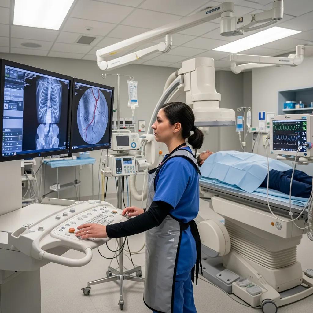
How Does Real-Time X-Ray Imaging Support Diagnosis and Intervention?
Real-time X-ray imaging through fluoroscopy significantly enhances diagnostic accuracy and treatment efficacy. This technique allows healthcare providers to visualize the anatomy and function of various organs as they operate, providing immediate insights that can influence clinical decisions. For instance, during gastrointestinal studies, fluoroscopy can reveal abnormalities in the digestive tract, enabling timely interventions. Additionally, the enhanced visualization of anatomy supports interventional radiology procedures, where precise navigation is crucial for successful outcomes.
What Clinical Applications Rely on Fluoroscopy Techniques?
Fluoroscopy techniques are essential in various clinical applications, including:
- Gastrointestinal Studies: Used to assess conditions such as ulcers, blockages, and motility disorders.
- Orthopedic Applications: Assists in the placement of screws and other hardware during surgeries.
- Cardiovascular Interventions: Guides catheter placements and stent insertions in real-time.
These applications highlight the versatility of fluoroscopy in addressing diverse medical needs.
How Is the Fluoroscopy Imaging Procedure Performed?
The fluoroscopy imaging procedure involves several key steps to ensure accurate results and patient safety.
- Preparation Steps for Patients: Patients may be required to fast for a specific period before the procedure, depending on the area being examined.
- Equipment Used in Fluoroscopy: The procedure typically involves an X-ray machine and a fluoroscope, which captures real-time images.
- Role of the Radiologist: A trained radiologist oversees the procedure, ensuring that images are captured correctly and that the patient is comfortable throughout.
It is essential to book an appointment with a qualified provider, such as X-Ray Docs, to ensure a smooth and professional experience.
What Are the Step-by-Step Fluoroscopy Procedure Guidelines?
The following guidelines outline the typical steps involved in a fluoroscopy procedure:
- Patient Positioning: The patient is positioned appropriately based on the area of interest.
- Contrast Agent Administration: A contrast agent may be administered to enhance the visibility of specific structures.
- Post-Procedure Care: After the imaging, patients are monitored for any immediate reactions to the contrast agent and provided with aftercare instructions.
These steps are crucial for obtaining high-quality images and ensuring patient safety.
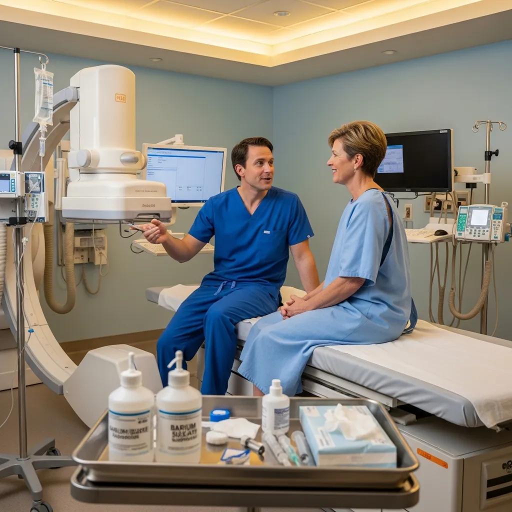
How Are Contrast Agents Used to Enhance Fluoroscopy Images?
Contrast agents play a vital role in fluoroscopy by improving the visibility of internal structures. These agents, which can be barium-based or iodine-based, help delineate organs and blood vessels, making it easier to identify abnormalities.
Research further highlights the specific role of barium sulfate in enhancing gastrointestinal imaging.
Barium Sulfate in Fluoroscopy: GI Imaging & Contrast
Barium sulfate is commonly used to radiographically examine the intestines, and improve visualization by opacifying areas of interest fluoroscopically. Typically, this is done by either infusing a water-soluble contrast or using barium. Barium sulfate can be given orally or rectally, and was initially commonly used in the beginning of the twentieth century; substituting for a prior m
Barium sulfate deposition in the gastrointestinal tract: review of the literature, DJ Zaccarini, 2022
| Contrast Agent | Type | Use |
|---|---|---|
| Barium Sulfate | Barium-based | Gastrointestinal studies |
| Iohexol | Iodine-based | Vascular imaging |
| Iopamidol | Iodine-based | Urological studies |
The choice of contrast agent depends on the specific imaging requirements and the patient’s medical history. Understanding the different types of contrast agents can help patients prepare for their procedures.
Despite their benefits, the use of fluoroscopic contrast media, particularly for GI and GU studies, has faced challenges related to availability and regulatory requirements.
Fluoroscopic Contrast Media: GI/GU Challenges & Safety
One of the significant challenges facing radiologists who perform and interpret studies of the gastrointestinal and genitourinary systems have been periodic interruptions in the availability of barium and iodinated contrast media specially formulated for gastrointestinal (GI) and genitourinary (GU) studies. These interruptions are due to the US Food and Drug Administration’s recent requirement for more stringent documentation of the safety and efficacy of contrast media and the consolidation among contrast manufacturers. This article reviews the current status of fluoroscopic contrast media, and provides suggestions and recommendations for the optimal and alternative use of contrast media formulations.
Contrast media for fluoroscopic examinations of the GI and GU tracts: current challenges and recommendations, MP Federle, 2017
What Are the Key Fluoroscopy Safety Guidelines and Radiation Protocols?
Safety is paramount in fluoroscopy, and several guidelines are in place to minimize radiation exposure.
- Radiation Exposure Limits: Healthcare providers adhere to strict protocols to limit the amount of radiation a patient receives during the procedure.
- Patient Monitoring: Continuous monitoring ensures that any adverse reactions to contrast agents are promptly addressed.
- Emergency Protocols: Facilities are equipped with emergency protocols to manage any unexpected complications.
These safety measures are designed to protect patients while ensuring effective imaging.
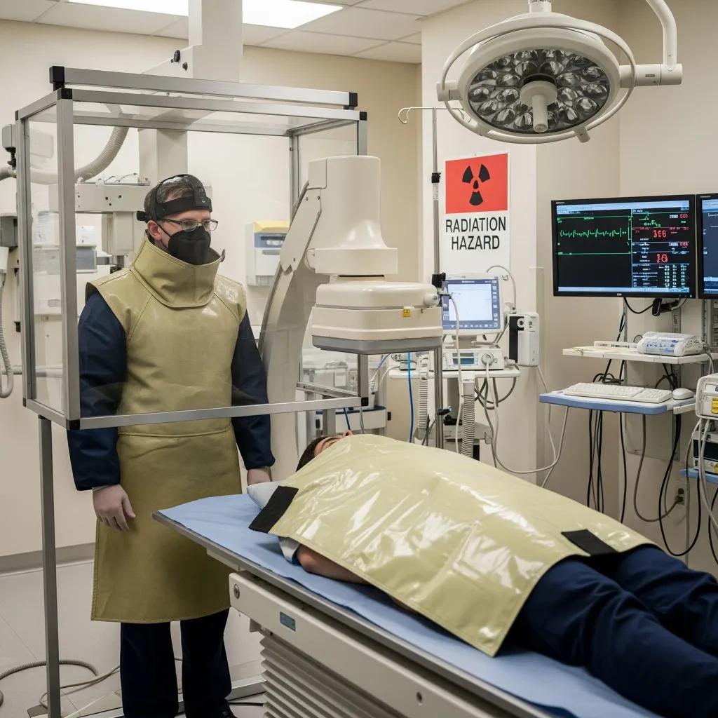
How Is Radiation Exposure Minimized During Fluoroscopy?
Minimizing radiation exposure during fluoroscopy is critical for patient safety.
- Use of Lead Shields: Protective lead shields are employed to cover areas not being imaged, reducing unnecessary exposure.
- Limiting Exposure Time: The duration of exposure is kept to a minimum, with real-time imaging allowing for quick assessments.
- Distance from Radiation Source: Maintaining an appropriate distance from the radiation source further reduces exposure levels.
These strategies collectively contribute to a safer imaging environment.
What Are the Recommended Patient Preparation and Safety Measures?
Proper patient preparation is essential for a successful fluoroscopy procedure.
- Dietary Restrictions: Patients may need to avoid certain foods or drinks before the procedure to ensure clear imaging.
- Clothing Considerations: Loose-fitting clothing is recommended, and patients may be asked to change into a gown.
- Communication with Healthcare Providers: Patients should inform their healthcare providers about any allergies or medical conditions that may affect the procedure.
By following these preparation measures, patients can help ensure a smooth and effective fluoroscopy experience.