Spine MRI vs CT Scan: Which Imaging Is Best for Diagnosing Back Pain?
When it comes to diagnosing back pain, choosing the right imaging technique is crucial for effective treatment. Two of the most common modalities are spine MRI and CT scans, each offering unique advantages and limitations. This article will explore the key differences between these imaging techniques, their specific applications, and how they can help in diagnosing various spinal conditions. Understanding these differences can empower patients to make informed decisions about their healthcare. We will also discuss when to choose each imaging type, the preparation required for each procedure, and how to book these services at X-Ray Docs, a leading provider of advanced imaging modalities in South Africa.
What Are the Key Differences Between Spine MRI and CT Scan?
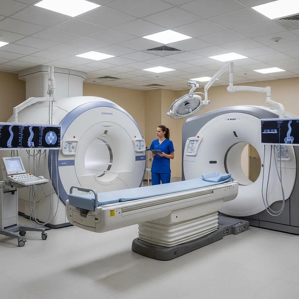
Spine MRI and CT scans are both essential tools in the evaluation of back pain, but they differ significantly in their technology and applications. MRI uses powerful magnets and radio waves to create detailed images of soft tissues, making it particularly effective for visualizing spinal discs, nerves, and muscles. In contrast, CT scans utilize X-rays to produce cross-sectional images, which are excellent for assessing bone structures and detecting fractures.
Cost is another factor to consider; generally, MRI scans tend to be more expensive than CT scans due to the advanced technology involved. However, the choice between the two often depends on the specific clinical scenario and the type of information needed by the healthcare provider.
How Do MRI and CT Scan Techniques Differ in Spinal Imaging?
The techniques used in MRI and CT scans are fundamentally different, leading to variations in the types of images produced. MRI employs a magnetic field and radiofrequency pulses to generate images, which can take longer to acquire but provide superior detail of soft tissues. This makes MRI the preferred choice for diagnosing conditions such as herniated discs and spinal stenosis.
On the other hand, CT scans are quicker and can be performed in emergency situations where time is of the essence. They are particularly useful for evaluating acute injuries and providing detailed images of bone structures, which is critical in trauma cases.
What Are the Advantages and Limitations of MRI and CT for Back Pain?
Both MRI and CT scans have their respective advantages and limitations when it comes to diagnosing back pain. Below is a comparison of these imaging modalities:
| Imaging Modality | Advantages | Limitations |
|---|---|---|
| MRI | Excellent for soft tissue evaluation, no radiation exposure | Longer scan times, higher cost |
| CT Scan | Quick imaging, superior for bone detail, less expensive | Limited soft tissue visualization, involves radiation |
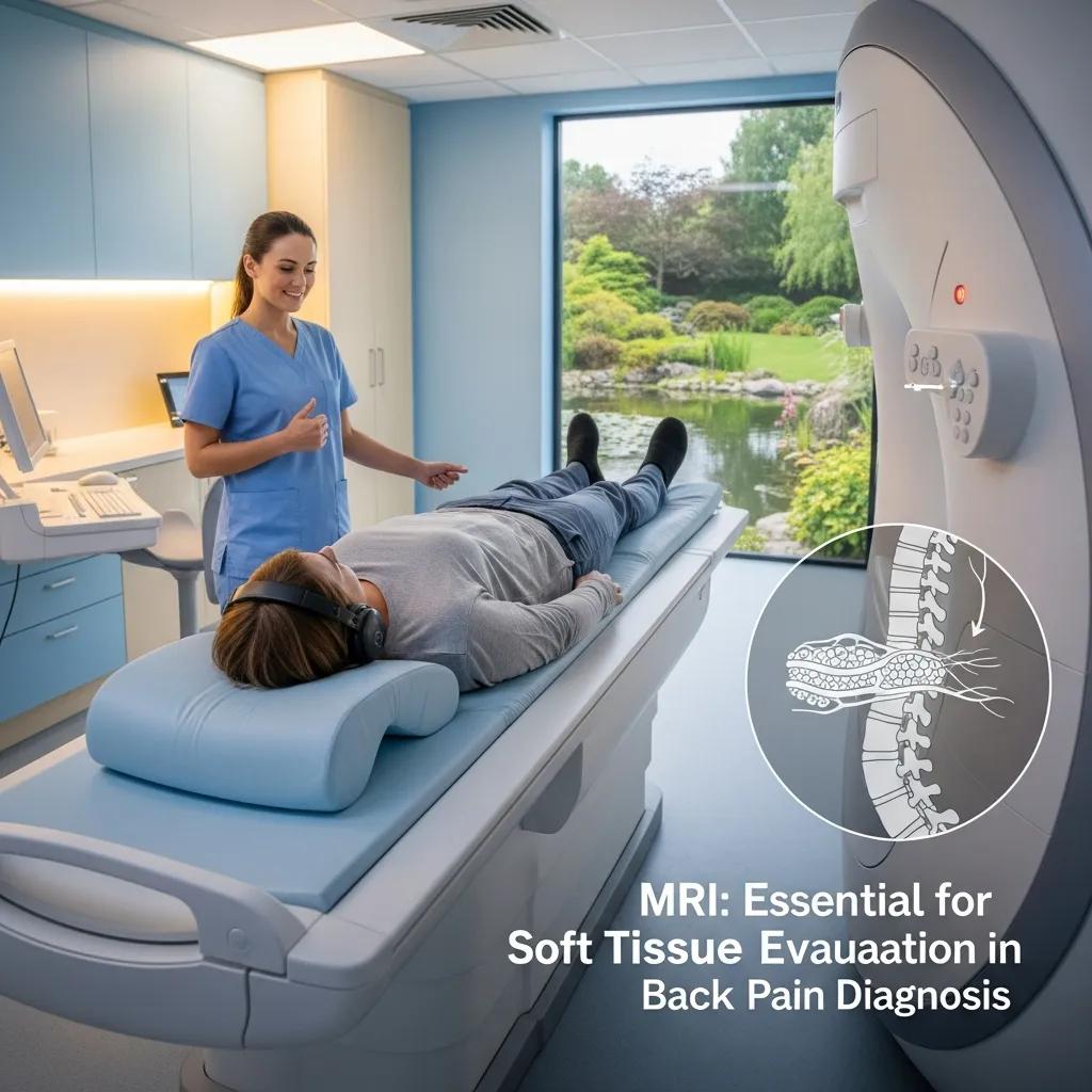
MRI is particularly advantageous for evaluating soft tissue injuries, while CT scans excel in providing detailed images of bone structures. Understanding these differences can help patients and healthcare providers make informed decisions about which imaging technique to use based on the specific clinical scenario.
When Should You Choose MRI or CT Scan for Back Pain Diagnosis?
The choice between MRI and CT scans for back pain diagnosis often depends on the clinical context. MRI is typically recommended for patients with suspected soft tissue injuries, such as herniated discs or spinal tumors. It is also the preferred method for evaluating chronic back pain when the underlying cause is unclear.
Adhering to established guidelines is crucial to ensure that imaging is performed only when clinically indicated, optimizing patient care and resource utilization.
Guidelines for Low Back Pain Imaging
Background Context The problem of imaging patients with low back pain (LBP) when it is not indicated is well recognized. The converse is also possible, although rarely considered.
Imaging for low back pain: is clinical use consistent with guidelines?
A systematic review and meta-analysis, HJ Jenkins, 2018
Conversely, CT scans are often chosen in emergency settings, particularly for assessing acute injuries, fractures, or when rapid diagnosis is necessary. Patients with a history of metal implants or claustrophobia may also be better suited for CT scans, as MRI can be challenging in these cases.
Which Spinal Conditions Are Better Detected by MRI?
MRI is particularly effective in diagnosing various spinal conditions, including:
- Herniated Discs: MRI provides detailed images of the spinal discs and surrounding soft tissues, making it easier to identify herniations.
- Spinal Stenosis: This condition, characterized by narrowing of the spinal canal, is best visualized through MRI.
- Tumors and Infections: MRI can detect tumors and infections in the spine that may not be visible on CT scans.
These conditions often require precise imaging to guide treatment decisions, making MRI an invaluable tool in spinal diagnostics.
When Is CT Scan Preferred for Spinal Injuries and Bone Detail?
CT scans are preferred in specific scenarios, particularly when detailed images of bone structures are required. They are especially useful for:
- Bone Fractures: CT scans can quickly identify fractures and assess their severity.
- Acute Injuries: In emergency situations, CT scans provide rapid imaging, which is crucial for timely intervention.
- Detailed Bone Imaging: CT is superior for visualizing complex bone anatomy, making it ideal for surgical planning.
In these cases, the speed and detail provided by CT scans can significantly impact patient outcomes.
How to Prepare for Spine MRI and CT Scan Procedures?

Preparation for spine MRI and CT scans is essential to ensure accurate results and patient safety. Here are the general preparation steps for each procedure:
What Are the Preparation Steps for a Spine MRI Scan?
Preparing for a spine MRI scan typically involves the following steps:
- Clothing Restrictions: Patients should wear comfortable clothing without metal fasteners, as metal can interfere with the MRI machine.
- Medication Considerations: Inform the healthcare provider about any medications being taken, especially those that may affect the scan.
How Should Patients Prepare for a Spine CT Scan?
Preparation for a spine CT scan may include:
- Dietary Restrictions: Patients may be advised to avoid food and drink for a few hours before the scan, especially if contrast material is used.
- Hydration Considerations: Staying hydrated is important, but patients should follow specific instructions regarding fluid intake prior to the scan.
Following these preparation guidelines can help ensure a smooth imaging process and accurate results.
How to Book Spine MRI and CT Scan Services at X-Ray Docs?
Booking spine MRI and CT scan services at X-Ray Docs is a straightforward process designed to accommodate patient needs. Here’s how to schedule an appointment:
What Is the Process for Scheduling Spine Imaging at X-Ray Docs?
Patients can schedule spine imaging services at X-Ray Docs through the following steps:
- Online Booking Options: Visit the X-Ray Docs website to book an appointment online.
- Phone Booking Process: Alternatively, patients can call to schedule their imaging services directly.
What Makes X-Ray Docs a Trusted Provider for Spine Imaging?
X-Ray Docs is recognized for its commitment to quality and patient care. Key factors that contribute to its reputation include:
- Expertise of Radiologists: The team consists of highly trained radiologists specializing in advanced imaging techniques.
- State-of-the-Art Technology: X-Ray Docs utilizes the latest imaging technology to ensure accurate diagnoses and effective treatment planning.
These attributes make X-Ray Docs a trusted choice for patients seeking spine imaging services in South Africa.