MRI Scan Overview: How It Works and What to Expect
Magnetic Resonance Imaging (MRI) is a non-invasive medical imaging technique that provides detailed images of the organs and tissues within the body. It utilizes powerful magnets and radio waves to create high-resolution images, allowing healthcare professionals to diagnose various conditions effectively. This article will explore the intricacies of MRI scans, including how they work, what to expect during the procedure, and essential preparation guidelines. Many patients may feel anxious about undergoing an MRI, but understanding the process can alleviate concerns and enhance the overall experience. We will cover the technology behind MRI, the components of an MRI machine, patient preparation, safety guidelines, and what happens during and after the scan.
What Is an MRI Scan and How Does Magnetic Resonance Imaging Work?
An MRI scan is a diagnostic imaging technique that uses magnetic fields and radio waves to generate detailed images of the body’s internal structures. The process works by aligning the protons in the body’s hydrogen atoms with a strong magnetic field. When radio waves are applied, these protons emit signals that are captured and transformed into images by a computer. This method is particularly beneficial for visualizing soft tissues, making it invaluable in diagnosing conditions related to the brain, spinal cord, muscles, and joints. The non-invasive nature of MRI scans allows for comprehensive assessments without the need for ionizing radiation, making it a safer alternative to other imaging techniques.
How Does MRI Technology Use Magnetic Fields and Radio Waves?
MRI technology relies on the principles of nuclear magnetic resonance (NMR). The strong magnetic field generated by the MRI machine causes the hydrogen nuclei in the body to align with the field. When radiofrequency pulses are applied, these nuclei are temporarily knocked out of alignment. As they return to their original state, they release energy in the form of radio waves, which are detected by the MRI machine. This process allows for the creation of detailed images of the body’s internal structures. The ability to manipulate magnetic fields and radio waves is what makes MRI a powerful tool in medical imaging, providing high-resolution images that can reveal abnormalities not visible through other imaging methods.
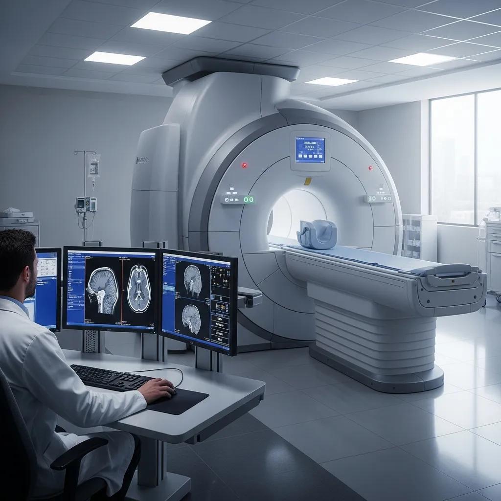
What Are the Key Components of an MRI Machine?

An MRI machine consists of several key components that work together to produce images. These include:
- Magnet: The most critical part of the MRI machine, the magnet generates a strong magnetic field that aligns the protons in the body.
- Gradient Coils: These coils create varying magnetic fields that allow for spatial encoding of the signals, enabling the production of detailed images.
- Radiofrequency Coils: These coils transmit radiofrequency pulses to the body and receive the signals emitted by the protons as they return to alignment.
- Computer System: The computer processes the signals received from the radiofrequency coils and constructs the images displayed on the monitor.
Understanding these components helps to appreciate the complexity and sophistication of MRI technology, which is essential for accurate diagnostics.
How Should You Prepare for an MRI Scan?

Preparing for an MRI scan involves several important steps to ensure the procedure runs smoothly. Patients should inform their healthcare provider about any medical conditions, allergies, or previous surgeries, especially if they have metal implants or devices. Additionally, patients may be advised to wear comfortable clothing without metal fasteners and to remove any jewelry or accessories before the scan.
What Are the Patient Preparation Guidelines for MRI Scans?
Here are some essential guidelines for patients preparing for an MRI scan:
- Inform Your Doctor: Disclose any medical history, especially regarding metal implants or devices.
- Wear Comfortable Clothing: Opt for loose-fitting clothes without metal components.
- Remove Accessories: Take off jewelry, watches, and any other metallic items before the scan.
Following these guidelines can help ensure a successful MRI experience.
Are There Specific Instructions for MRI Contrast Agents?
In some cases, a contrast agent may be used to enhance the quality of the images obtained during an MRI scan. Patients should follow specific instructions regarding the use of contrast agents, which may include:
- Hydration: Drink plenty of water before the scan to help flush the contrast agent from the body afterward.
- Allergy Information: Inform the healthcare provider of any allergies, particularly to iodine or gadolinium, which are common components of contrast agents.
- Pre-Scan Instructions: Follow any dietary restrictions or medication guidelines provided by the healthcare team.
These precautions help minimize risks and improve the effectiveness of the MRI scan.
What Are the Safety Guidelines and Contraindications for MRI Scans?
Safety is a paramount concern when undergoing an MRI scan. The following guidelines should be observed:
- Metal Implants: Patients with certain metal implants, such as pacemakers or cochlear implants, may not be eligible for an MRI due to the strong magnetic field.
- Pregnancy: Pregnant women should discuss the necessity of the scan with their healthcare provider, as the effects of MRI on the fetus are not fully understood.
- Claustrophobia: Patients who experience anxiety in confined spaces should inform their healthcare provider, as sedation may be necessary.
These safety measures ensure that the MRI procedure is conducted without risk to the patient.
Is MRI Safe and Who Should Avoid It?
MRI is generally considered safe for most individuals. However, certain populations should exercise caution or avoid the procedure altogether. Patients with the following conditions should consult their healthcare provider before undergoing an MRI:
- Metal Implants: Individuals with ferromagnetic implants may be at risk due to the strong magnetic field.
- Pregnant Women: While MRI does not use ionizing radiation, its effects on fetal development are still being studied.
- Severe Anxiety: Those with claustrophobia or anxiety disorders may require additional support or alternative imaging methods.
Understanding these considerations can help patients make informed decisions regarding their imaging options.
What Precautions Do Radiologists Take During MRI Procedures?
Radiologists take several precautions to ensure patient safety and comfort during MRI procedures. These include:
- Screening: Thoroughly screening patients for contraindications and medical history before the scan.
- Monitoring: Continuously monitoring patients during the procedure to address any discomfort or concerns.
- Emergency Protocols: Having emergency protocols in place in case of adverse reactions to contrast agents or other unexpected events.
These measures help to create a safe and supportive environment for patients undergoing MRI scans.
What to Expect During and After Your MRI Scan in South Africa
During an MRI scan, patients can expect to lie still on a padded table that slides into the MRI machine. The procedure typically lasts between 30 to 60 minutes, depending on the area being scanned. Patients may hear loud tapping or thumping noises during the scan, which is normal. Earplugs or headphones may be provided to help minimize discomfort from the noise.
After the scan, patients can resume normal activities unless otherwise instructed. If a contrast agent was used, drinking plenty of water is recommended to help flush it from the system.
What Is the Step-by-Step MRI Scan Procedure?
The MRI scan procedure generally follows these steps:
- Preparation: Patients change into a gown and remove any metal objects.
- Positioning: The patient is positioned on the MRI table, and the area of interest is aligned with the machine.
- Scanning: The MRI machine begins to take images, and patients must remain still during this time.
- Post-Scan: Once the scan is complete, patients can leave and await results from their healthcare provider.
This structured approach ensures that the MRI scan is conducted efficiently and effectively.
How Are MRI Results Interpreted and Delivered?
After the MRI scan, a radiologist interprets the images and prepares a report detailing the findings. This report is then sent to the referring physician, who will discuss the results with the patient. Depending on the findings, further tests or treatments may be recommended. Patients can typically expect to receive their results within a few days to a week, depending on the facility’s protocols.
In South Africa, X-Ray Docs is a trusted provider of MRI scans, specializing in advanced imaging technology and patient-centered care. For those seeking to book an MRI appointment, X-Ray Docs offers a streamlined process to ensure a smooth experience.