Ultrasound vs X-Ray: Choosing the Best Medical Imaging Scan for Your Condition
When it comes to medical imaging, choosing the right scan can significantly impact diagnosis and treatment. Ultrasound and X-ray are two common imaging techniques, each with unique mechanisms and applications. This article will explore the differences between ultrasound and X-ray, helping you understand which scan is best suited for your specific condition. By the end, you will have a clearer understanding of the benefits and limitations of each imaging method, as well as guidance on when to choose one over the other.
What Are Ultrasound and X-Ray Imaging Techniques?
Ultrasound and X-ray imaging are essential tools in modern medicine, providing critical insights into various health conditions. Ultrasound imaging uses high-frequency sound waves to create images of organs and structures inside the body. This non-invasive technique is particularly useful for examining soft tissues, such as the heart, liver, and developing fetus during pregnancy. In contrast, X-ray imaging employs ionizing radiation to produce images of the body’s internal structures, primarily bones. This method is widely used for diagnosing fractures, infections, and other skeletal issues.
How Does Ultrasound Imaging Work?
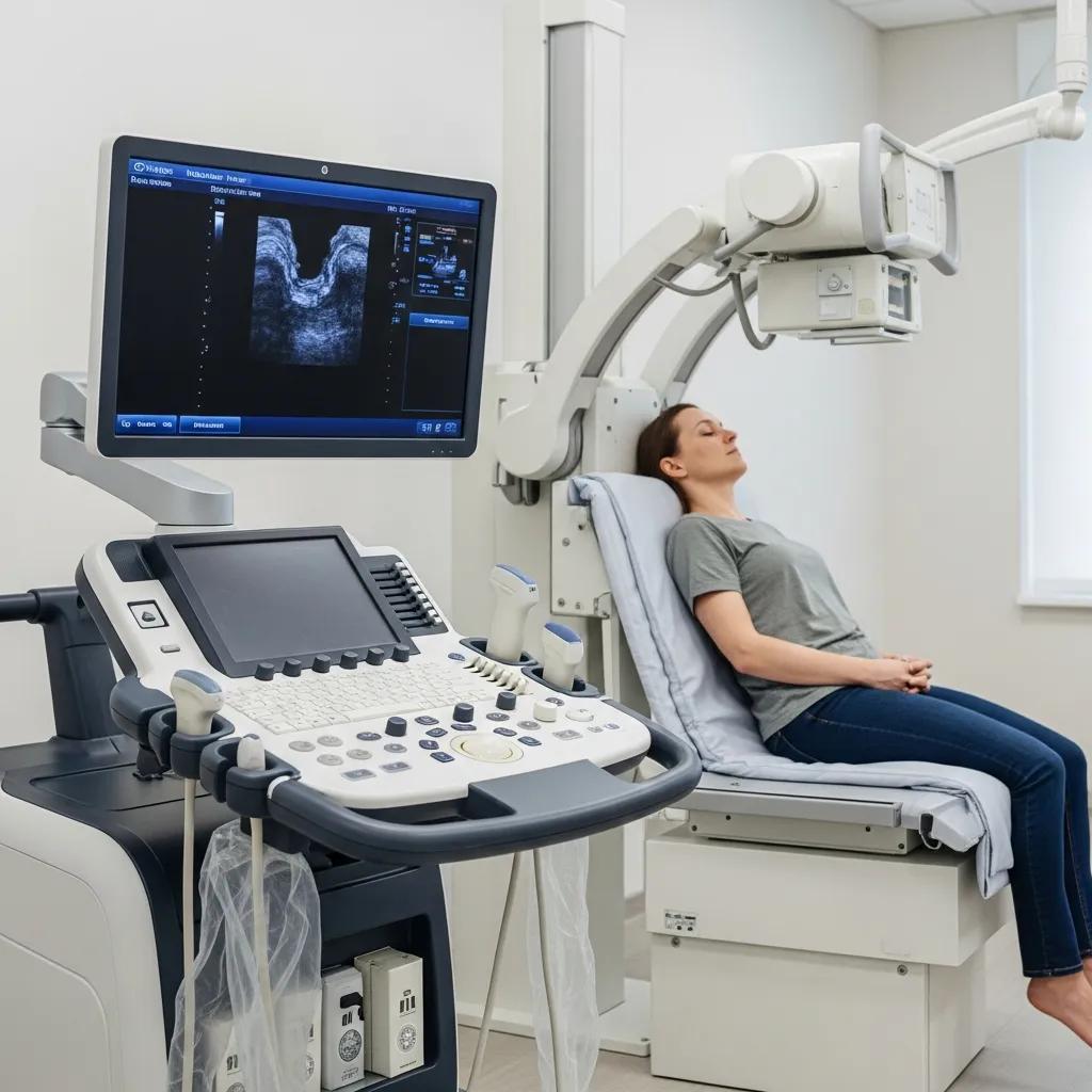
Ultrasound imaging operates by emitting sound waves through a transducer, which then captures the echoes that bounce back from internal structures. The resulting data is processed to create real-time images displayed on a monitor. This technique is non-invasive and does not involve radiation, making it a safe option for various diagnostic purposes. Ultrasound is particularly effective in monitoring pregnancy, assessing abdominal organs, and guiding certain medical procedures.
What Is X-Ray Imaging and How Is It Performed?
X-ray imaging involves the use of ionizing radiation to create images of the body’s internal structures. During the procedure, a patient is positioned between an X-ray machine and a digital detector. The machine emits radiation that passes through the body, capturing images of bones and other dense tissues. X-rays are commonly used to diagnose fractures, dental issues, and chest conditions. Safety measures, such as lead aprons, are often employed to minimize radiation exposure to patients.
While X-ray imaging is invaluable for diagnosis, it’s important to consider the associated radiation exposure and the necessity for clinical justification and dose optimization, especially for vulnerable populations.
X-Ray Radiation Exposure: Clinical Justification and Dose Optimization
Radiation doses associated with dual-energy X-ray absorptiometry are very low. However, as with any X-ray imaging technique, each particular examination must always be clinically justified. When an examination is justified, the emphasis must be on dose optimisation of imaging protocols. Dose optimisation is more important for paediatric examinations because children are more vulnerable to radiation than adults. Methods based on multi-detector CT (MDCT) are associated with higher radiation doses. New 3D volumetric hip and spine quantitative computed tomography (QCT) techniques and high-resolution MDCT for evaluation of bone structure deliver doses to patients from 1 to 3 mSv. Low-dose protocols are needed to reduce radiation exposure from these methods and minimise associated health risks.
Radiation exposure in X-ray-based imaging techniques used in osteoporosis, J Damilakis, 2010
What Are the Key Differences Between Ultrasound and X-Ray Scans?
Understanding the key differences between ultrasound and X-ray scans is crucial for making informed decisions about medical imaging. Each technique has its own safety profiles, diagnostic capabilities, and limitations.
How Do Ultrasound and X-Ray Differ in Safety and Radiation Exposure?

One of the most significant differences between ultrasound and X-ray imaging is the safety profile. Ultrasound is completely radiation-free, making it a preferred choice for pregnant women and children. In contrast, X-ray imaging involves exposure to ionizing radiation, which carries a small risk of potential harm, particularly with repeated exposure. However, the benefits of X-ray imaging often outweigh the risks, especially when diagnosing serious conditions.
Reinforcing the benefits of ultrasound, particularly its safety, research underscores its value as a non-ionizing and rapid imaging modality.
Musculoskeletal Ultrasound: Non-Ionizing, Safe, and Rapid Imaging
Musculoskeletal ultrasound (MSK-US) is a non-ionizing imaging modality, which is relatively inexpensive, portable, safe and rapid [1–4].
The accuracy of diagnostic ultrasound imaging for musculoskeletal soft tissue pathology of the extremities: a comprehensive review of the literature, KJ Young, 2015
What Are the Diagnostic Accuracy and Limitations of Each Scan?
Both ultrasound and X-ray have their strengths and limitations in diagnostic accuracy. Ultrasound excels in visualizing soft tissues and fluid-filled structures, making it ideal for assessing conditions like cysts, tumors, and organ abnormalities. However, it may not provide clear images of dense tissues like bones. On the other hand, X-ray imaging is highly effective for diagnosing bone fractures and certain lung conditions but may not be as reliable for soft tissue evaluation. Understanding these differences can help guide the choice of imaging technique based on the specific medical condition.
When Should You Choose Ultrasound or X-Ray for Diagnosis?
Choosing between ultrasound and X-ray depends on the medical condition being evaluated. Each imaging technique is suited for different diagnostic purposes.
Which Medical Conditions Are Best Diagnosed with Ultrasound?
Ultrasound is particularly effective for diagnosing several medical conditions, including:
- Pregnancy Monitoring: Ultrasound is the standard method for monitoring fetal development and assessing pregnancy-related complications.
- Abdominal Issues: It is commonly used to evaluate conditions affecting the liver, gallbladder, kidneys, and pancreas.
- Soft Tissue Evaluation: Ultrasound is ideal for assessing soft tissue injuries, such as tears in ligaments or muscles.
When Is X-Ray Preferred for Imaging?
X-ray imaging is preferred for diagnosing conditions such as:
- Bone Fractures: X-rays are the go-to method for identifying fractures and dislocations.
- Dental Issues: Dentists frequently use X-rays to detect cavities, impacted teeth, and other dental problems.
- Chest Imaging: X-rays are essential for diagnosing pneumonia, tumors, and other lung conditions.
How to Book Your Ultrasound or X-Ray Scan at X-Ray Docs
Booking an imaging scan is a straightforward process, especially at X-Ray Docs, a South African diagnostic imaging provider specializing in X-ray and ultrasound services.
What Is the Step-by-Step Appointment Booking Process?

To book your ultrasound or X-ray scan at X-Ray Docs, follow these steps:
- Visit the Website: Go to the X-Ray Docs website to access the appointment booking section.
- Select Service and Date: Choose the type of imaging service you need and select a convenient date and time for your appointment.
- Provide Personal Information: Fill out the required personal information, including your name, contact details, and any relevant medical history.
What Should Patients Know Before Their Imaging Scan?
Before attending your imaging appointment, consider the following preparation tips:
- Clothing and Dietary Restrictions: Wear comfortable clothing and follow any dietary restrictions if required for your specific scan.
- Necessary Information: Bring any relevant medical records or referrals from your healthcare provider.
- Potential Risks: Be aware of any potential risks associated with the imaging technique, especially if you have allergies or specific health conditions.
| Imaging Technique | Safety Profile | Diagnostic Strengths | Limitations |
|---|---|---|---|
| Ultrasound | Radiation-free | Excellent for soft tissues | Limited for dense tissues |
| X-Ray | Ionizing radiation | Superior for bone imaging | Less effective for soft tissues |
This comparison highlights the distinct safety profiles and diagnostic strengths of ultrasound and X-ray imaging, guiding patients in making informed decisions about their healthcare.
In summary, both ultrasound and X-ray imaging play vital roles in medical diagnostics, each with unique advantages and limitations. Understanding these differences can help patients and healthcare providers make informed decisions about the most appropriate imaging technique for specific medical conditions.